This article will elucidate all the mysteries surrounding the thoracic vertebrae and will describe both their typical and atypical features. The thoracic vertebrae are located in the middle section of the vertebral column, specifically inferior to the cervical vertebrae and superior to the lumbar vertebrae.
T1 (1st Thoracic Vertebra) The T1 vertebra is the first (uppermost) of the twelve (12) thoracic vertebrae that make up the central and largest section of the spinal column between the lumbar vertebrae below and the cervical vertebrae above. While larger than the C7 vertebra above it, the T1 is the smallest of the thoracic vertebrae.
T1 (1st Thoracic Vertebra) The T1 vertebra is the first (uppermost) of the twelve (12) thoracic vertebrae that make up the central and largest section of the spinal column between the lumbar vertebrae below and the cervical vertebrae above. While larger than the C7 vertebra above it, the T1 is the smallest of the thoracic vertebrae.
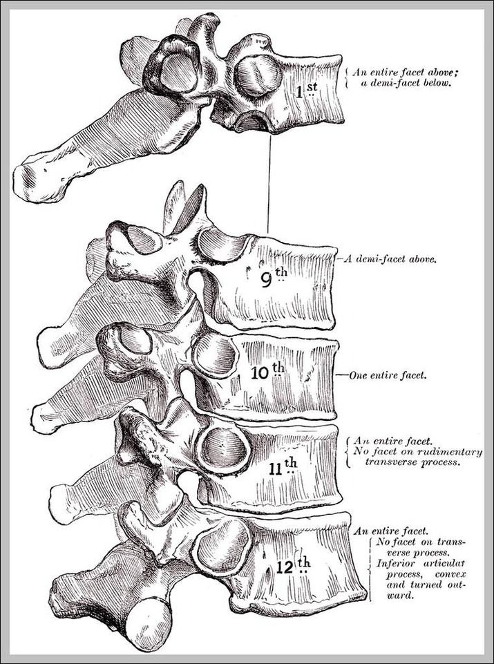
Thoracic Vertebra Image
Posted inDiagrams