Capsular Ligament Image

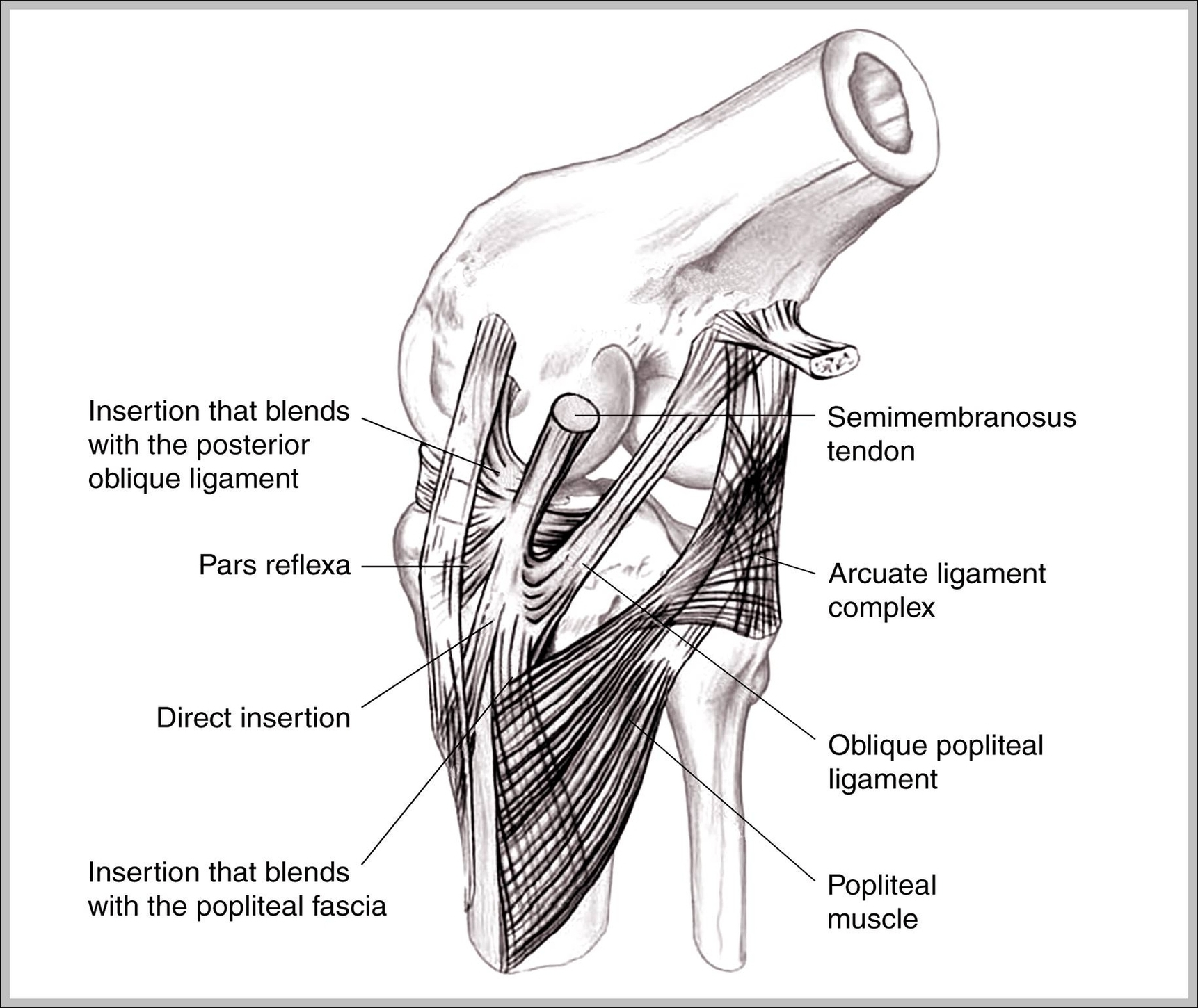
In joint: Joint ligaments Capsular ligaments are simply thickenings of the fibrous capsule itself that take the form of either elongated bands or triangles, the fibres of which radiate from a small area of one articulating bone to a line upon its mating fellow.
Recognition of the normal magnetic resonance (MR) imaging appearances of the capsular ligaments of the knee is of great importance. These ligaments contribute to stability of the knee joint and are frequently injured.

Capsular Ligament Image Diagram - Capsular Ligament Image Chart - Human anatomy diagrams and charts explained. This anatomy system diagram depicts Capsular Ligament Image with parts and labels. Best diagram to help learn about health, human body and medicine.

Capsular Ligament Image

Medial Collateral Ligament Image

The medial collateral ligament ( MCL) of the knee is a flat, triangular band on its medial aspect that resists valgus forces. It forms part of the medial capsuloligamentous complex of the knee . The medial collateral ligament measures 8-10 cm in length and has superficial and deep portions 4.
MCL sprains are graded 1, 2 or 3 depending on severity. The medial collateral ligament (or MCL for short) connects the thigh bone (femur) to the shin bone (tibia) on the inside of your knee. It provides stability to your knee, preventing it from moving sideways.

Medial Collateral Ligament Image Diagram - Medial Collateral Ligament Image Chart - Human anatomy diagrams and charts explained. This anatomy system diagram depicts Medial Collateral Ligament Image with parts and labels. Best diagram to help learn about health, human body and medicine.

Medial Collateral Ligament Image

Inguinal Ligament Exercises Image

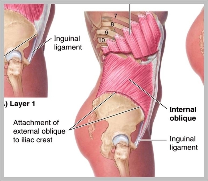
Inguinal Ligament Workout. Swimming is another very effective cardiovascular exercise. The water provides for a medium that minimizes the impact of external forces on the inguinal ligament injury as well as leads to the strengthening of the entire body structure and thereby the ligaments as well.
Your inguinal ligaments are fibrous bands of tissue flanking your inner groin on each side. Usually taut, the ligaments help support your pelvic and abdominal areas.

Inguinal Ligament Exercises Image Diagram - Inguinal Ligament Exercises Image Chart - Human anatomy diagrams and charts explained. This anatomy system diagram depicts Inguinal Ligament Exercises Image with parts and labels. Best diagram to help learn about health, human body and medicine.

Inguinal Ligament Exercises Image

Fibular Collateral Ligament Image

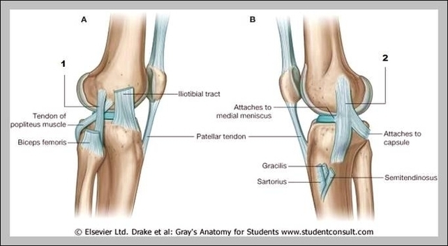
Medically reviewed by Healthline’s Medical Network on April 2, 2015. The fibular collateral ligament is one of the ligaments that make up the knee joint. Ligaments are bands of fibrous, durable tissue that connect and strengthen joints. They can be likened to rubber bands.
Stress radiographs might show increased opening 5. An arcuate sign/fracture can be seen in case of an avulsion injury of the fibular tip 2-4. MRI allows for the localization of the injury and injury grading. Fibular collateral ligament injuries can be best depicted in coronal and axial views 2.
The lateral collateral ligament (LCL) is a thin band of connective tissue that runs along the outside of the knee. It connects the femur to the fibula and stabilizes the knee, bracing it from unusual impact.

Fibular Collateral Ligament Image Diagram - Fibular Collateral Ligament Image Chart - Human anatomy diagrams and charts explained. This anatomy system diagram depicts Fibular Collateral Ligament Image with parts and labels. Best diagram to help learn about health, human body and medicine.

Fibular Collateral Ligament Image