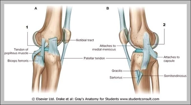
Fibular Collateral Ligament Image

Medically reviewed by Healthline’s Medical Network on April 2, 2015. The fibular collateral ligament is one of the ligaments that make up the knee joint. Ligaments are bands of fibrous, durable tissue that connect and strengthen joints. They can be likened to rubber bands.
Stress radiographs might show increased opening 5. An arcuate sign/fracture can be seen in case of an avulsion injury of the fibular tip 2-4. MRI allows for the localization of the injury and injury grading. Fibular collateral ligament injuries can be best depicted in coronal and axial views 2.
The lateral collateral ligament (LCL) is a thin band of connective tissue that runs along the outside of the knee. It connects the femur to the fibula and stabilizes the knee, bracing it from unusual impact.

Fibular Collateral Ligament Image Diagram - Fibular Collateral Ligament Image Chart - Human anatomy diagrams and charts explained. This anatomy system diagram depicts Fibular Collateral Ligament Image with parts and labels. Best diagram to help learn about health, human body and medicine.

Fibular Collateral Ligament Image