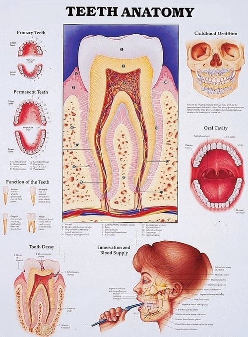
These nerve tissues and blood vessels are extending from the jaw bone to the pulp through a small hole at the root tip. Tooth Anatomy: Every tooth consists of three parts: the crown, neck, and root. Also, every tooth made of several layers: the enamel, dentin, cementum, and pulp.
The tooth is made of several layers of varying density and hardness: the enamel, dentin, cementum, and pulp. The enamel is the outer layer of teeth that covers the crown. Also, it protects the tooth against external damaging forces. Tooth enamel is the most resistant and hardest tissue in the human body.
The tooth is made of several layers of varying density and hardness: the enamel, dentin, cementum, and pulp. The enamel is the outer layer of teeth that covers the crown. Also, it protects the tooth against external damaging forces. Tooth enamel is the most resistant and hardest tissue in the human body.
Teeth anatomy explained
Posted inDiagrams
Teeth anatomy explained
Post navigation
Previous Post
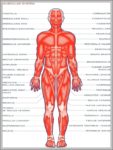 Major Function Of Muscular System Image