4,924 ovaries stock photos and images available, or search for ovaries icon or polycystic ovaries to find more great stock photos and pictures. A good ultrasound image can identify a…
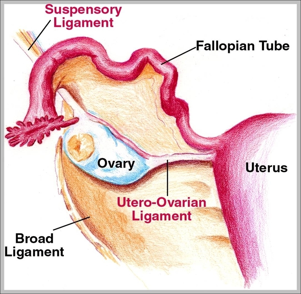
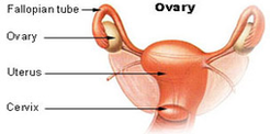
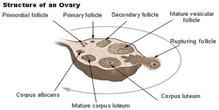
Structure of the Ovary. The ovaries are almond-shaped structures located on either side of the uterus, and closely related to several anatomical structures in the pelvic region. Each ovary has…
Structure of the Ovary. The ovaries are almond-shaped structures located on either side of the uterus, and closely related to several anatomical structures in the pelvic region. Each ovary has…