
Nerves Of The Lower Leg Image
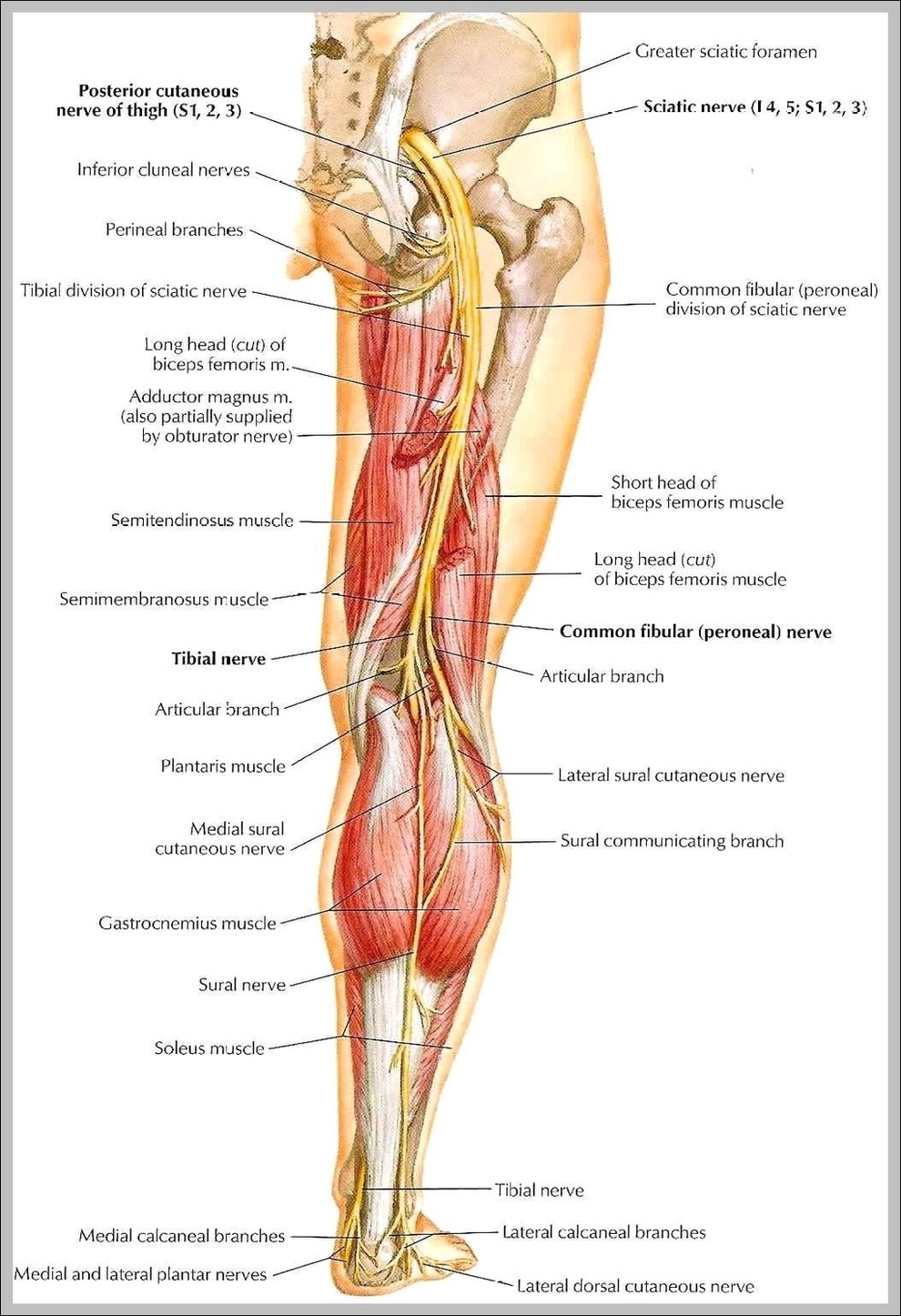
There are two main nerves in the leg: the femoral nerve serves the front and the sciatic nerve controls the back of the leg. The nerves of the leg can have many nerve roots, and when pain or discomfort is felt in these areas, it usually indicates a compressed or pinched nerve.
The lower leg is a major anatomical part of the skeletal system. Together with the upper leg, it forms the lower extremity. It lies between the knee and the ankle, while the upper leg lies between the hip and the knee. The lower leg contains two major long bones, the tibia and the fibula, which are both very strong skeletal structures.
The main muscle in this area of the leg is the gastrocnemius, which gives the calf a bulging muscular appearance. Some nerves of the sacral plexus innervate this area, namely the superficial fibular nerve, the deep fibular nerve and the tibial nerve.