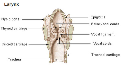
General Anatomy of the Larynx – Larynx Anatomy. The walls of the larynx are made up of cartilage, ligaments, membranes, muscles, and respiratory mucosa (or mucous membrane). There are nine (9) laryngeal cartilages, three (3) paired and three (3) single single. Together, they form a supportive skeletal framework.
Click on Label for the labeled model. Back to Respiratory System. The larynx is a guarded air passageway between the pharynx and the trachea. It is formed by 9 supportive cartilages, intrinsic and extrinsic muscles and a mucous membrane lining.
The extrinsic laryngeal muscles move the larynx as a whole. They consist of the suprahyoid muscles that elevate the hyoid bone and the larynx during swallowing and vocalization, and the infrahyoid muscles that depress the hyoid bone and the larynx.
larynx diagram
Posted inDiagrams
Larynx diagram
Post navigation
Previous Post
Next Post
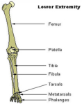
Lower extremity diagram