Common iliac vein. The common iliac vein is formed by the unification of the internal and external iliac veins. The external iliac vein drains the lower limb, and the internal iliac vein drains the gluteal region and pelvic viscera. The unification of the two common iliac veins forms the inferior vena cava.
Iliac Vein Compression Syndrome. Left common iliac vein stenosis frequently occurs where the vein crosses beneath the right common iliac artery. Chronic, repetitive compression at this site causes fibrosis of the vein, with synechiae and spurs that result in stenosis or even occlusion of the lumen.
Iliac Vein Compression Syndrome. Left common iliac vein stenosis frequently occurs where the vein crosses beneath the right common iliac artery. Chronic, repetitive compression at this site causes fibrosis of the vein, with synechiae and spurs that result in stenosis or even occlusion of the lumen.
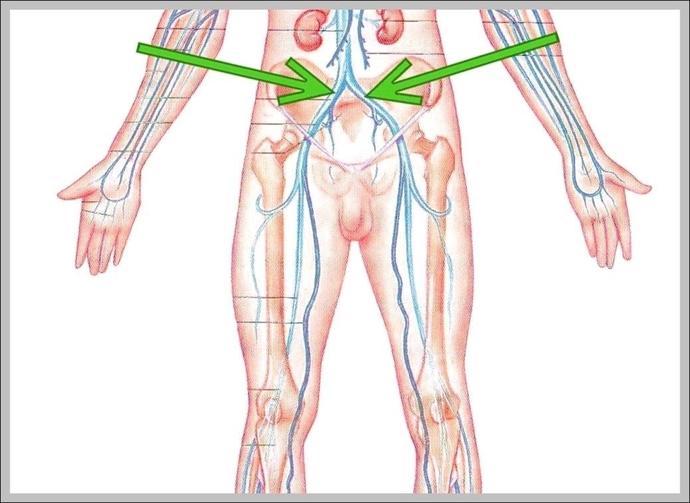
Common Iliac Vein Image
Posted inDiagrams
Common Iliac Vein Image
Post navigation
Previous Post
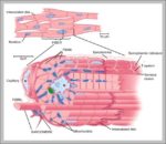 Heart Muscle Diagram Image
Next Post
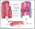
Artery And Vein Image