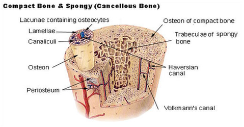
Structure of Bone Tissue. There are two types of bone tissue: compact and spongy. The names imply that the two types differ in density, or how tightly the tissue is packed together. There are three types of cells that contribute to bone homeostasis.
Figure 6.3.3 â Anatomy of a Flat Bone: This cross-section of a flat bone shows the spongy bone (diploë) covered on either side by a layer of compact bone. Osseous tissue is a connective tissue and like all connective tissues contains relatively few cells and large amounts of extracellular matrix.
Four types of cells are found within bone tissue. Osteogenic cells are undifferentiated and develop into osteoblasts. When osteoblasts get trapped within the calcified matrix, their structure and function changes, and they become osteocytes.
bone tissue diagram
Posted inDiagrams
Bone tissue diagram
Post navigation
Previous Post
Next Post
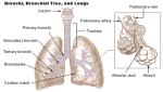
Bronchi lungs diagram