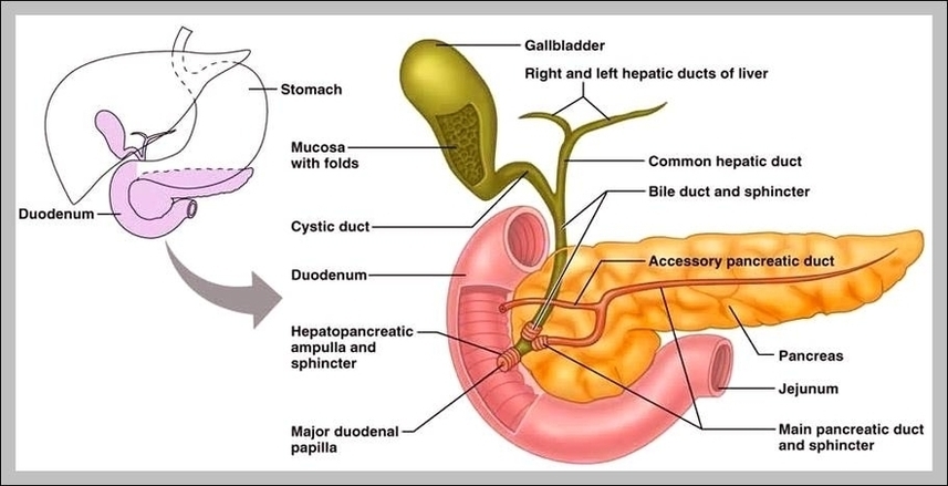
Though small in size, the gallbladder plays an important role in our digestion of food. The gallbladder holds bile produced in the liver until it is needed for digesting fatty…
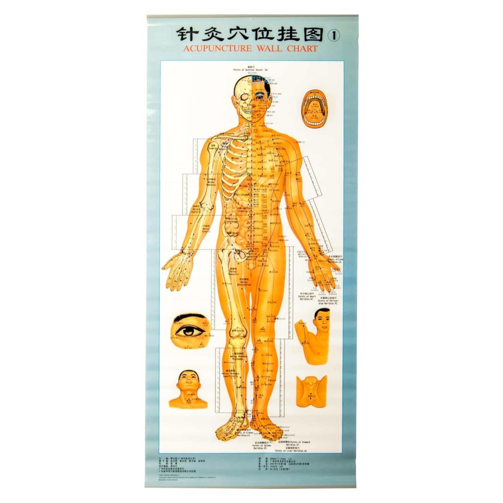
Extraordinary points are non-meridian points note contained within the 12 main meridians. This chart contains 64 extraordinary points with point location and indications for each point. 60 points mapped out…
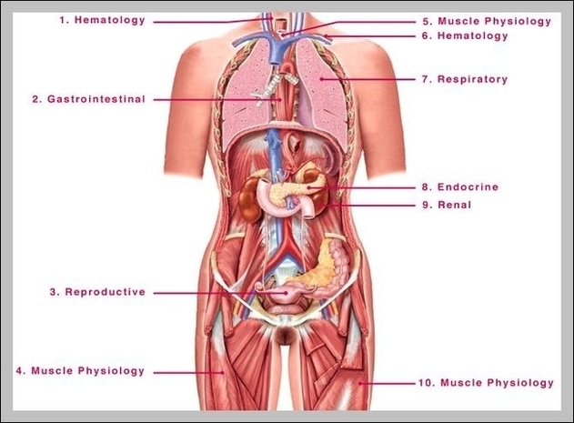
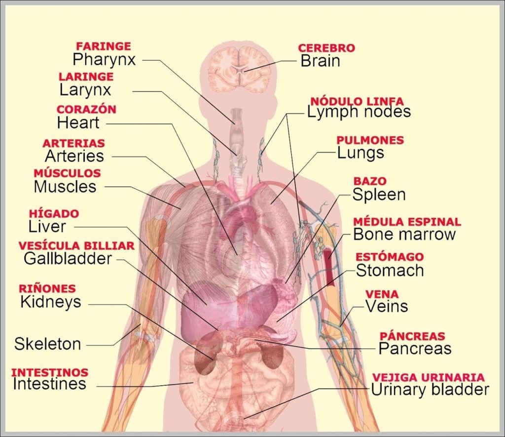
7,751 organs of the human body diagram stock illustrations and vector graphics available royalty-free, or start a new search to explore more great stock images and vector art. internal organs…
407,393 human internal organ stock photos and images available, or search for human internal organ icons or human internal organ illustrations to find more great stock photos and pictures. Organs,…
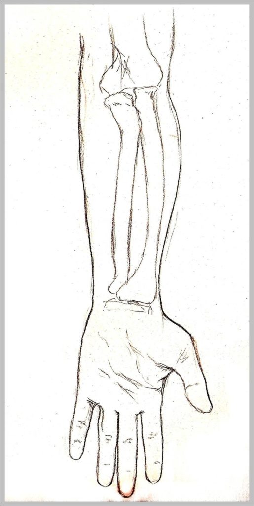
Pronation and supination of the hand: Anatomy and biomechanics Proper functioning of the hand relies on its capacity to rotate and point the palm upward (i.e. supination) or downward (i.e.…
WebMD's Knee Anatomy Page provides a detailed image and definition of the knee and its parts including ligaments, bones, and muscles. Skip to main content X-rays of the knee are…
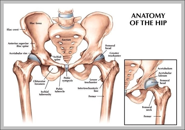
4,902 hip anatomy stock photos and images available, or search for human hip anatomy or hip anatomy illustration to find more great stock photos and pictures. General Hip Anatomy. The…
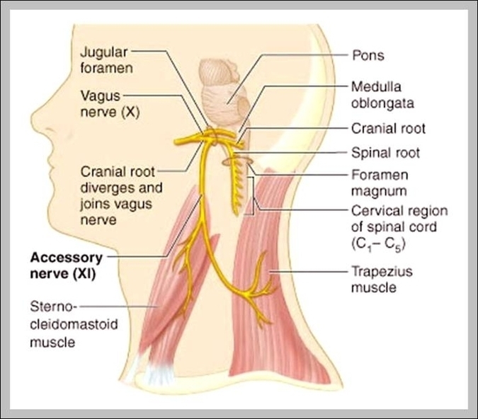
The cranial nerves are a set of twelve nerves that originate in the brain. Each has a different function for sense or movement. The functions of the cranial nerves are…
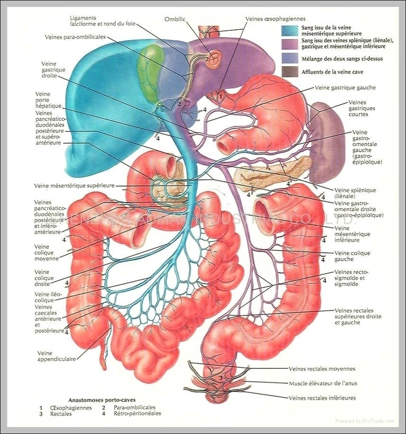
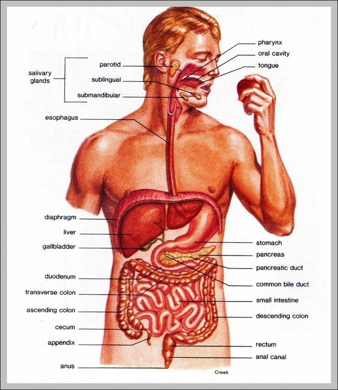
51,216 "human digestive system" stock photos, vectors, and illustrations are available royalty-free. The human digestive system consists of the gastrointestinal tract plus the accessory organs of digestion. Anatomy of human…
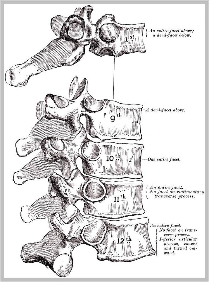
This article will elucidate all the mysteries surrounding the thoracic vertebrae and will describe both their typical and atypical features. The thoracic vertebrae are located in the middle section of…
Endocrine System Diagram The endocrine system consists of glands that are found all over the body, which help you to produce hormones. The endocrine system is a collection of glands…
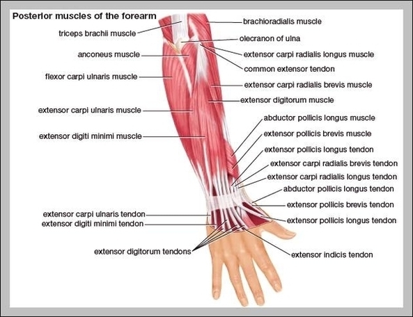
The muscles of the forearm can be divided into two groups: anterior (flexors) and posterior (extensors). Both the flexors and extensors are further divided into superficial and deep layers. The…
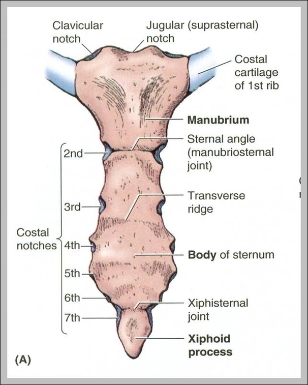
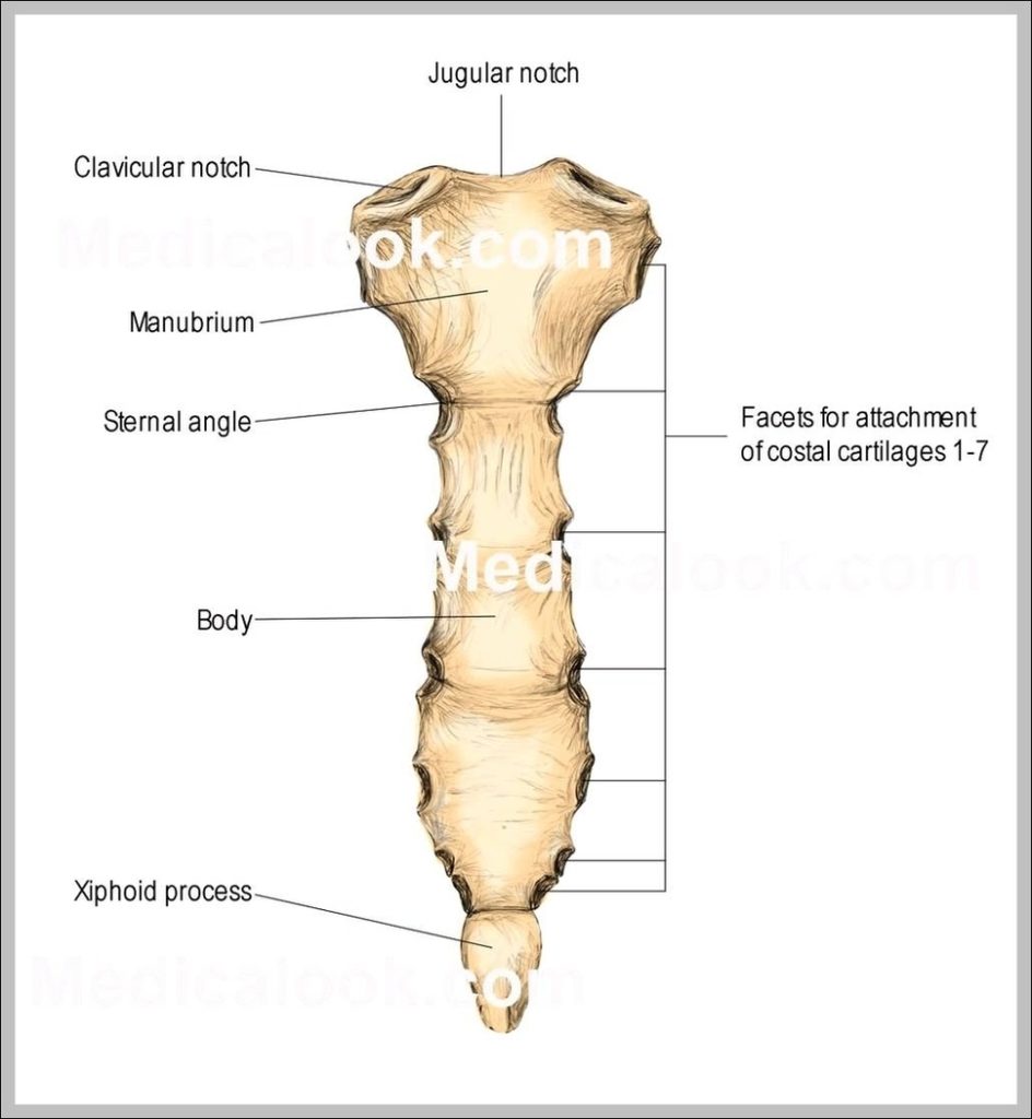
The sternum, commonly known as the breastbone, is a long, narrow flat bone that serves as the keystone of the rib cage and stabilizes the thoracic skeleton. The oblique sternum…
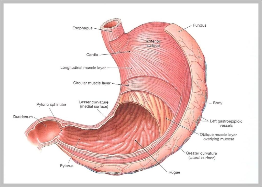
Anatomy of the Stomach. The stomach is an organ of the digestive system. It is an expanded section of the digestive tube between the esophagus and small intestine. Its characteristic…
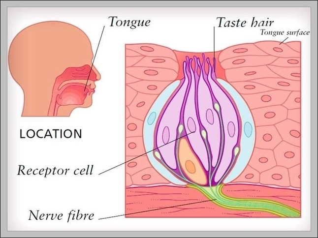
2,294 taste buds stock photos and images available, or search for tongue taste buds or taste buds macro to find more great stock photos and pictures. Human tongue, Lingual papillae…
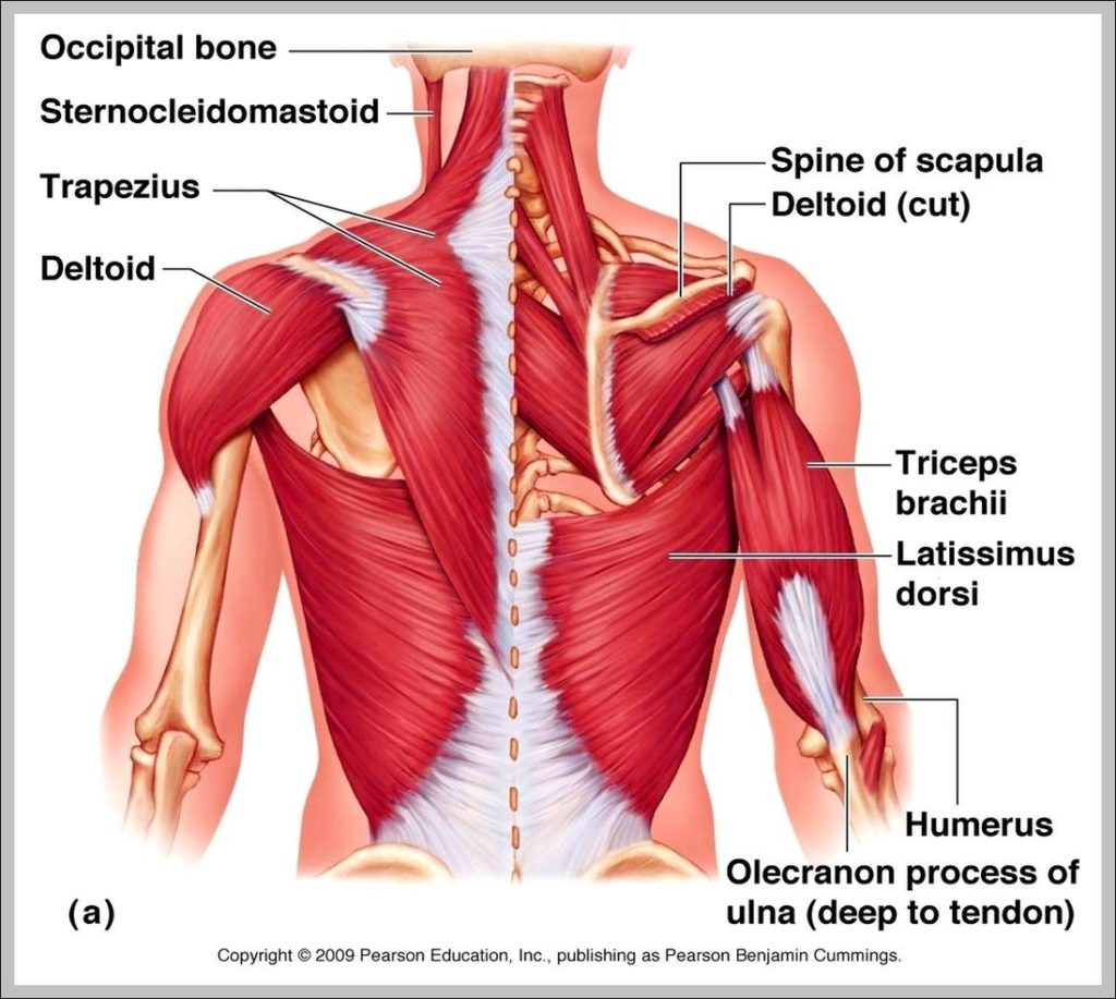
6,953 human upper body anatomy stock photos, vectors, and illustrations are available royalty-free. See human upper body anatomy stock video clips 128,412 muscle anatomy stock photos, vectors, and illustrations are…
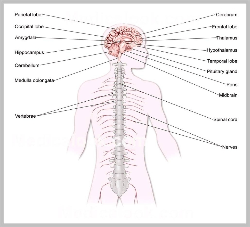
77,434 human nervous system stock photos and images available, or search for human nervous system anatomy or human nervous system brain to find more great stock photos and pictures. Human…