The cranial nerves are a set of twelve nerves that originate in the brain. Each has a different function for sense or movement. The functions of the cranial nerves are sensory, motor, or both:
Magnetic resonance imaging (MRI) is considered the gold standard in the study of the cranial nerves. Computed tomography (CT) allows, usually, an indirect view of the nerve and is useful to demonstrate the intraosseous segments of cranial nerves, the foramina through which they exit skull base and their pathologic changes.
Magnetic resonance imaging (MRI) is considered the gold standard in the study of the cranial nerves. Computed tomography (CT) allows, usually, an indirect view of the nerve and is useful to demonstrate the intraosseous segments of cranial nerves, the foramina through which they exit skull base and their pathologic changes.
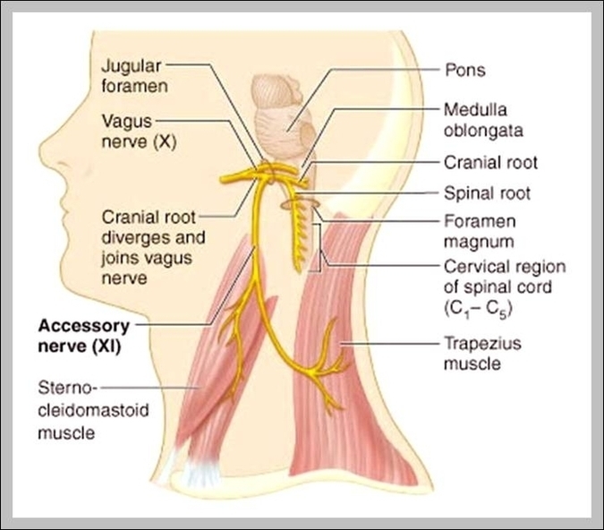
Cranial Nerve X Image
Posted inDiagrams
Cranial Nerve X Image
Post navigation
Previous Post
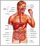 Pictures Of The Human Digestive System Image
Next Post
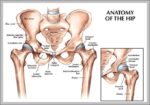
Hip Bone Anatomy Diagram Image