Medial and Lateral Pectoral Nerve Diagram

Medial and Lateral Pectoral Nerve Diagram

Medial pectoral nerve (C8-T1) pierces pectoralis minor to supply it and pectoralis major sternocostal head; lateral pectoral nerve (C5-C7) supplies clavicular head of pectoralis major. Both from brachial plexus.
Course of the Long Thoracic Nerve Diagram

Course of the Long Thoracic Nerve Diagram

The long thoracic nerve (C5-C7) descends superficially on serratus anterior, supplying it for scapular protraction/rotation. Injury causes winged scapula and difficulty raising arm overhead.
Recurrent Branch of Median Nerve Diagram

Recurrent Branch of Median Nerve Diagram

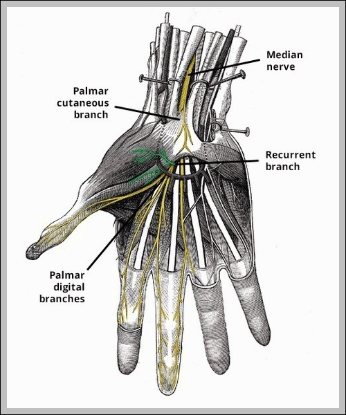
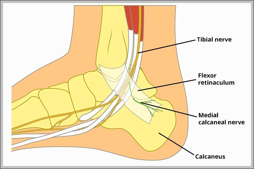
Recurrent branch of median nerve arises distal to carpal tunnel, loops around flexor pollicis brevis to supply thenar muscles (abductor pollicis brevis, opponens pollicis, superficial flexor pollicis brevis head). It’s…
Course of the Lacrimal Nerve Diagram

Course of the Lacrimal Nerve Diagram

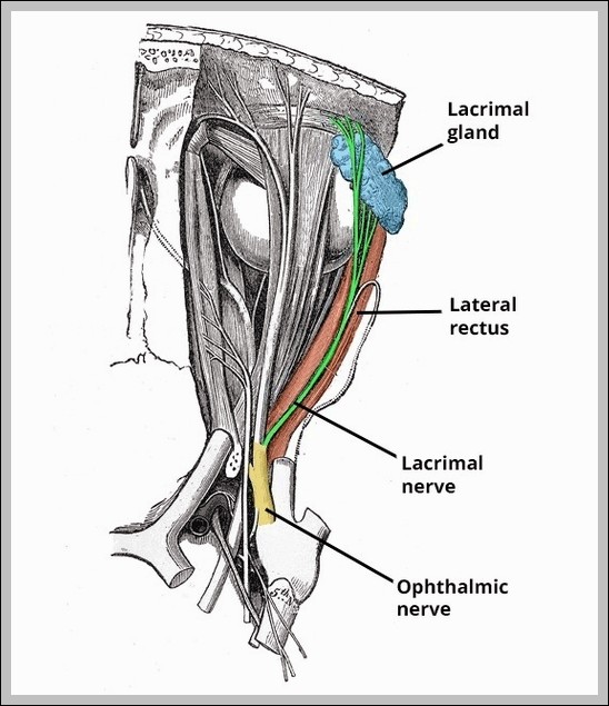
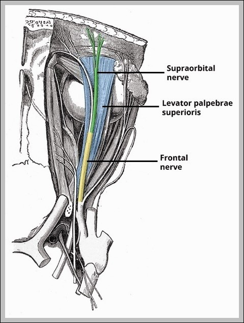
Lacrimal nerve (V1 branch) enters orbit via superior orbital fissure, runs superolateral, supplies lacrimal gland (parasympathetic hitchhike from facial via pterygopalatine ganglion), conjunctiva, upper eyelid skin. It communicates with zygomatic…
Zygomatic Nerve Course Branches Diagram

Zygomatic Nerve Course Branches Diagram

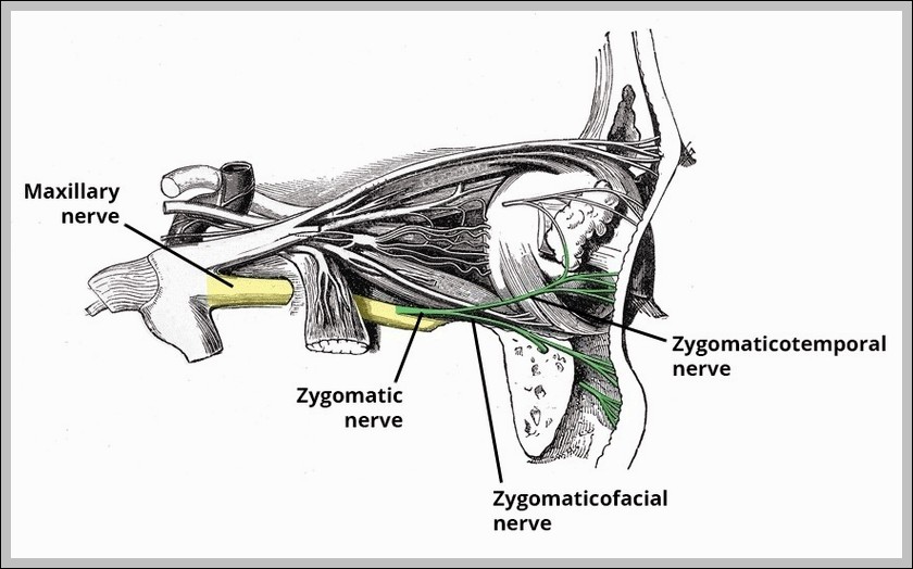
The zygomatic nerve (V2 branch) enters orbit via inferior orbital fissure, gives zygomaticotemporal (skin over temple) and zygomaticofacial (cheek) branches, and communicates with lacrimal nerve for parasympathetics.
Radial Nerve Palsy Wristdrop Diagram

Radial Nerve Palsy Wristdrop Diagram

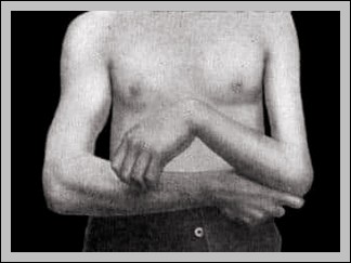
Radial nerve palsy causes wrist drop (loss of extensors), sensory loss dorsum hand/thumb-index-middle. Common from humeral shaft fracture (spiral groove) or Saturday night compression.
Anatomical Course of the Musculocutaneous Nerve Diagram

Anatomical Course of the Musculocutaneous Nerve Diagram

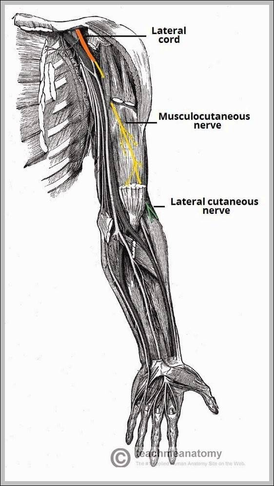
The musculocutaneous nerve (C5-C7, lateral cord) pierces coracobrachialis, runs between biceps/brachialis, supplies coracobrachialis, biceps, brachialis (elbow flexion/supination), then becomes lateral cutaneous nerve of forearm for lateral forearm skin.
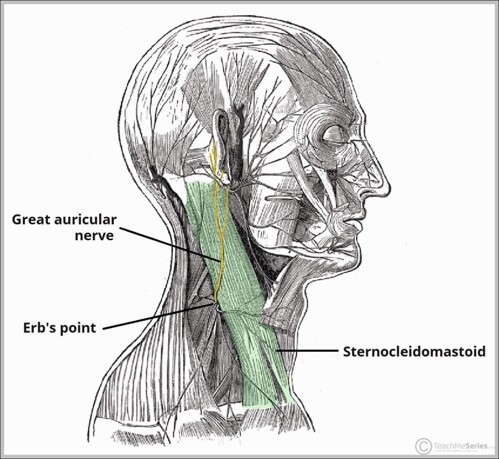
Course of the Great Auricular Nerve Diagram

Course of the Great Auricular Nerve Diagram

The great auricular nerve (C2-C3 from cervical plexus) ascends behind sternocleidomastoid, emerges at Erb’s point, and supplies skin over parotid, mastoid, auricle, and angle of mandible. It’s commonly blocked for…
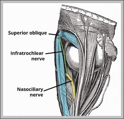
Infratrochlear Nerve Course Supply Diagram

Infratrochlear Nerve Course Supply Diagram

The infratrochlear nerve (terminal branch of nasociliary, V1) exits orbit below trochlea, supplies sensation to medial upper eyelid, caruncle, lacrimal sac, root of nose, and skin between eye and nose…
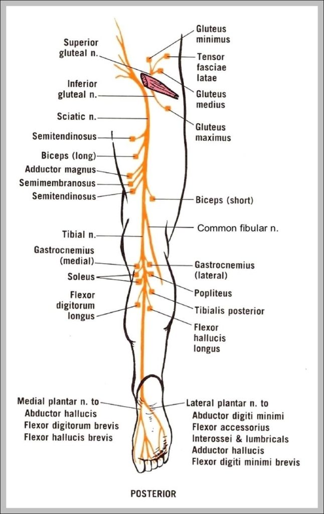
Sciatic Nerve Anatomy Diagram Image

Sciatic Nerve Anatomy Diagram Image

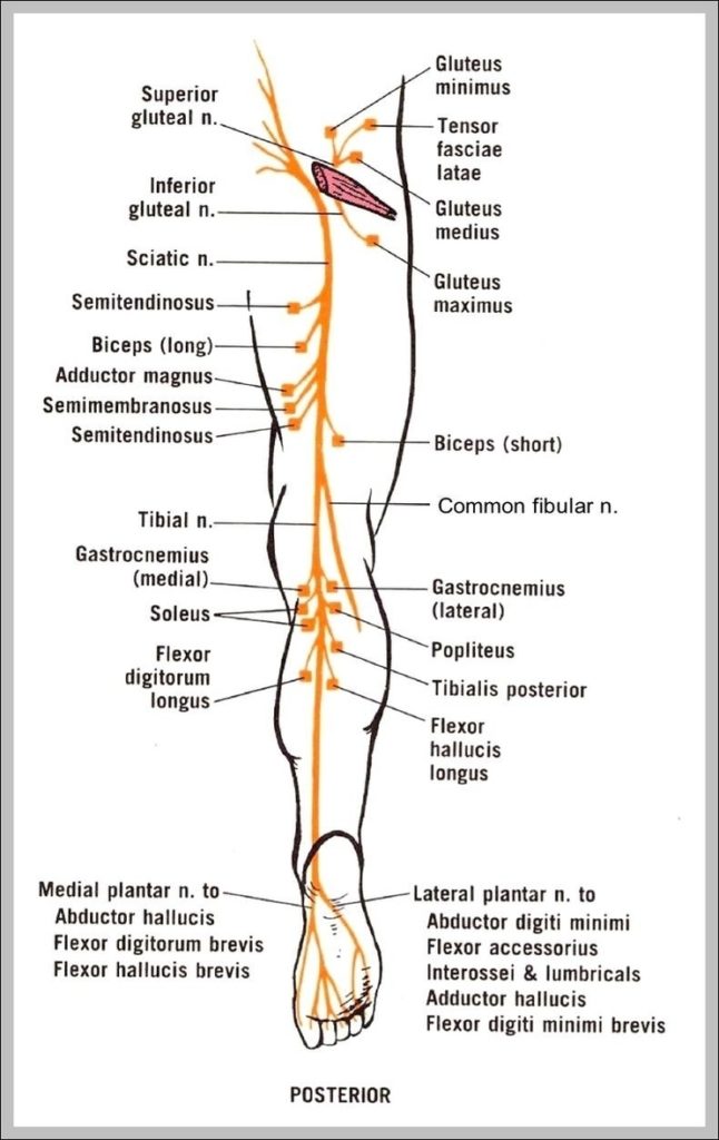
The sciatic nerve is the largest and longest spinal nerve in the human body. Extending from the lumbar and sacral plexuses in the lower back, the sciatic nerve runs through…
Cranial Nerve X Image

Cranial Nerve X Image

The cranial nerves are a set of twelve nerves that originate in the brain. Each has a different function for sense or movement. The functions of the cranial nerves are…