Skip to content
cropped anatomy systems Anatomy System - Human Body Anatomy diagram and chart images

Human Body Anatomy Diagrams

  • Diagrams
  • Organs
  • Bones
  • Disease
    • Cancer
  • Muscles
  • Cell
  • Health
    • Dental
    • Medical
    • Women
  • Best
  • Home
  • Medical
  • Anatomy of the Medial Calcaneal Nerve Diagram
Anatomy of the Medial Calcaneal Nerve Diagram
Posted inMedical

Anatomy of the Medial Calcaneal Nerve Diagram

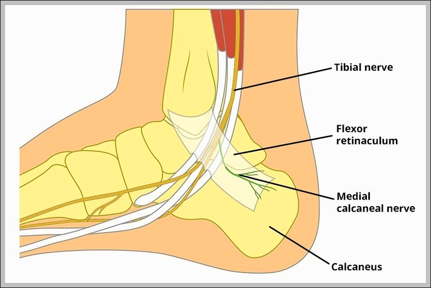
Medial calcaneal nerve (tibial branch) supplies sensation to medial heel skin, emerges proximal to tarsal tunnel. Entrapment causes medial heel pain mimicking plantar fasciitis.

Tags:
anatomycalcanealdiagrammedialnerve

Post navigation

Previous Post
Anatomical Course of the Musculocutaneous Nerve Diagram Anatomical Course of the Musculocutaneous Nerve Diagram
Next Post
Radial Nerve Palsy Wristdrop Diagram Radial Nerve Palsy Wristdrop Diagram
  • Overview of the Urinary Tract Kidney Ureter and Bladder Diagram
  • Structure of the Testes and Epididymis Diagram
  • Tooth
  • Descent of the Testis Embryology Diagram
  • Muscular Triangle of the Neck Diagram
Copyright 2026 — Anatomy System - Human Body Anatomy diagram and chart images. All rights reserved. Bloghash WordPress Theme
Scroll to Top