Human Respiratory System Visual

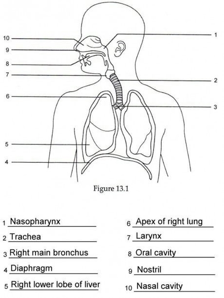
The Human Respiratory System

The human respiratory system is a complex network of organs and structures responsible for the vital process of respiration, which involves the exchange of gases between the air we breathe and our body’s cells.

Upper Respiratory System

The upper respiratory system comprises the nose, nasal cavities, and the paranasal sinuses. These structures provide airways for respiration. The pharynx, also part of the upper respiratory system, connects the nasal and oral cavities to the larynx and esophagus. The larynx and vocal cords allow us to breathe, talk, and sing.

Lower Respiratory System

The lower respiratory system consists of the larynx, trachea, bronchi, and the structures within the lungs, such as the bronchioles and alveolar ducts. The trachea is the main airway to the lungs, while the bronchi are passageways that facilitate the movement of air in and out of the lungs.

Lungs and Alveoli

The lungs, located in the thorax and protected by the bony and muscular thoracic cage, are responsible for gas exchange between the air we breathe and our bodies. The alveoli, microscopic air sacs within the lungs, are the site of this external respiration.

Diaphragm

The diaphragm, the main respiratory muscle, plays an essential role in the physical process of breathing. Under the control of the central nervous system, the diaphragm and the intercostal muscles of the chest wall generate a pumping action on the lung, expanding and contracting the internal space of the thorax.

Function of the Respiratory System

Through breathing, inhalation and exhalation, the respiratory system facilitates the exchange of gases between the air and the blood and between the blood and the body’s cells. The respiratory system also helps us to smell and create sound.

Conclusion

The human respiratory system is a marvel of biological engineering, efficiently facilitating the exchange of life-sustaining gases. Understanding its structure and function is crucial to comprehending how our bodies utilize oxygen and expel waste gases, maintaining the delicate balance necessary for life..

Human Respiratory System Visual Diagram - Human Respiratory System Visual Chart - Human anatomy diagrams and charts explained. This anatomy system diagram depicts Human Respiratory System Visual with parts and labels. Best diagram to help learn about health, human body and medicine.

Human Respiratory System Visual

A Skeleton With Labelshuman Skeleton Diagram With Labels Visual

The human skeleton, an intricate framework of bones and cartilage, provides essential support, protection, and mobility for our bodies. Comprising 206 bones in adults, the skeleton is a remarkable system that plays a crucial role in maintaining our physical form and enabling various functions.

## 1. Structure and Composition:
– The human skeleton consists of two main divisions: the axial skeleton and the appendicular skeleton.
– Axial Skeleton:
– Forms the central axis of the body.
– Includes the skull, vertebral column, and rib cage.
– The skull comprises the cranium (protecting the brain) and the facial bones.
– The vertebral column (or spine) consists of 33 vertebrae, providing flexibility and support.
– The rib cage safeguards vital organs like the heart and lungs.
– Appendicular Skeleton:
– Attaches to the axial skeleton.
– Includes the limbs, shoulder girdle, and pelvic girdle.
– The shoulder girdle connects the upper limbs to the axial skeleton.
– The pelvic girdle supports the lower limbs and protects reproductive organs.

## 2. Bone Types:
– Bones are classified into five types based on shape and function:
1. Long Bones:
– Longer than they are wide.
– Examples: femur (thigh bone), humerus (upper arm bone).
2. Short Bones:
– Cube-shaped.
– Provide stability and support.
– Examples: carpals (wrist bones), tarsals (ankle bones).
3. Flat Bones:
– Thin and flat.
– Protect internal organs and provide attachment points.
– Examples: scapula (shoulder blade), sternum (breastbone).
4. Irregular Bones:
– Complex shapes.
– Serve specific functions.
– Examples: vertebrae, facial bones.
5. Sesamoid Bones:
– Develop within tendons.
– Reduce friction and enhance mechanical advantage.
– Example: patella (kneecap).

## 3. Bone Tissue:
– Bones consist of compact bone (dense and strong) and spongy bone (trabecular bone with a porous structure).
– Osteocytes, embedded in the bone matrix, maintain bone health.
– Haversian canals contain blood vessels and nerves, ensuring nutrient supply.

## 4. Joints:
– Joints connect bones and allow movement.
– Types of joints:
– Fibrous Joints: Immovable (e.g., sutures in the skull).
– Cartilaginous Joints: Slightly movable (e.g., intervertebral discs).
– Synovial Joints: Freely movable (e.g., ball-and-socket, hinge joints).
– Examples: shoulder joint, hip joint, knee joint.

## 5. Bone Development and Growth:
– Ossification:
– Process of bone formation.
– Intramembranous ossification: Forms flat bones directly from mesenchymal tissue.
– Endochondral ossification: Converts cartilage into bone (long bones).
– Bone Remodeling:
– Ongoing process of resorption and deposition.
– Maintains bone strength and adapts to stress.

## 6. Common Bones:
– Skull Bones:
– Cranium (frontal, parietal, temporal, occipital, sphenoid, ethmoid).
– Facial bones (maxilla, mandible, zygomatic, nasal, etc.).
– Vertebral Column:
– Cervical (neck), thoracic (mid-back), lumbar (lower back), sacral, and coccygeal vertebrae.
– Upper Limb Bones:
– Humerus, radius, ulna, carpals, metacarpals, and phalanges.
– Lower Limb Bones:
– Femur, tibia, fibula, patella, tarsals, metatarsals, and phalanges.

## 7. Clinical Significance:
– Osteoporosis: Bone density loss.
– Fractures: Breaks in bones.
– Arthritis: Joint inflammation.
– Scoliosis: Abnormal spinal curvature.

In summary, the human skeleton is a marvel of engineering, providing both strength and flexibility. Its intricate design ensures our survival, movement, and protection throughout life..

A Skeleton With Labelshuman Skeleton Diagram With Labels Visual Diagram - A Skeleton With Labelshuman Skeleton Diagram With Labels Visual Chart - Human anatomy diagrams and charts explained. This anatomy system diagram depicts A Skeleton With Labelshuman Skeleton Diagram With Labels Visual with parts and labels. Best diagram to help learn about health, human body and medicine.

A Skeleton With Labelshuman Skeleton Diagram With Labels Visual

Human Body Muscles Visual

The human body is a complex system, and the muscles are a crucial part of it. Muscles are responsible for all movement in the body, from the beating of the heart to the lifting of a finger. Understanding the visual aspect of human body muscles can be facilitated through various online platforms and applications.

Zygote Body 3D Anatomy Online Visualizer is a free online 3D anatomy atlas that allows you to view, isolate, and learn about human anatomy structures. It provides a 3D model of the human body, which you can rotate, zoom, and manipulate to better understand the structure and function of different muscles. The platform also offers an opacity slider to reveal layers and a search box to select specific entities.

Another platform is Visible Body, which creates interactive anatomy and physiology products for the web and mobile devices. It offers a visual and interactive approach to learning about the human body, including the muscular system. It provides hundreds of animations and simulations, and you can test your knowledge with dynamic quizzes and flashcards.

The Human Anatomy Atlas 2024 app provides muscle and bone models that you can move. It allows you to learn about muscle actions, bone landmarks, attachments, innervations, and blood supply.

These platforms provide a comprehensive and interactive way to study the human body’s muscular system. They offer a variety of tools to help you visualize and understand the complexity and functionality of muscles. From the major muscle groups like the biceps and quadriceps to the intricate muscles of the face and hand, these platforms provide a detailed and interactive way to study and understand the human muscular system.

In conclusion, visualizing the human body muscles can significantly enhance our understanding of human anatomy. It provides a more intuitive and engaging way to learn about the structure and function of muscles. Whether you’re a student, a healthcare professional, or just someone interested in learning more about the human body, these platforms offer a wealth of information at your fingertips. Remember, a picture is worth a thousand words, and in the case of learning about human body muscles, a 3D visual can be priceless..

Human Body Muscles Visual Diagram - Human Body Muscles Visual Chart - Human anatomy diagrams and charts explained. This anatomy system diagram depicts Human Body Muscles Visual with parts and labels. Best diagram to help learn about health, human body and medicine.

Human Body Muscles Visual

Internal Structure Of Human Stomach Visual

The human stomach, a key organ in the digestive system, is a complex structure with several distinct regions and layers. It plays a crucial role in the digestion of food and absorption of nutrients.

Location and Structure

The stomach is located in the upper abdomen on the left side of the body. It is a J-shaped organ that spans the region between the cardiac and pyloric orifices of the gastrointestinal tract. The stomach’s convex lateral surface is known as the greater curvature, while the concave medial border is the lesser curvature.

Parts of the Stomach

The stomach comprises four major regions:
1. Cardia: The area around the opening where the esophagus connects to the stomach.
2. Fundus: The dome-shaped part located to the left of the cardia.
3. Body: The main, central region of the stomach.
4. Pylorus: The lower part of the stomach that connects to the duodenum.

Layers of the Stomach

The stomach wall consists of several layers:
1. Mucosa: The innermost layer, which produces enzymes and acids for digestion.
2. Submucosa: Contains connective tissue, blood vessels, lymph vessels, and nerve cells.
3. Muscularis Externa: The primary muscle of the stomach, responsible for churning and mixing food.
4. Serosa: The outermost layer, a membrane that covers the stomach.

Function of the Stomach

The stomach’s primary function is to digest food and send it to the small intestine. It temporarily stores food, contracts and relaxes to mix and break down food, and produces enzymes and other specialized cells to digest food. The stomach works in conjunction with the rest of the gastrointestinal tract to break down food and liquid, absorb nutrients and water, and expel waste products of digestion through the large intestine.

Blood Supply and Innervation

The stomach receives its blood supply mainly from the celiac trunk. Innervation is provided via the vagus nerves and the celiac plexus.

Microscopic Anatomy

The inner part of the stomach lining, the gastric mucosa, consists of an outer layer of column-shaped cells, a lamina propria, and a thin layer of smooth muscle called the muscularis mucosa. Beneath the mucosa lies the submucosa, consisting of fibrous connective tissue.

In conclusion, the human stomach is a complex organ with a detailed internal structure. Its various parts and layers work together to perform the essential function of digesting food and absorbing nutrients, making it a vital component of the human digestive system..

Internal Structure Of Human Stomach Visual Diagram - Internal Structure Of Human Stomach Visual Chart - Human anatomy diagrams and charts explained. This anatomy system diagram depicts Internal Structure Of Human Stomach Visual with parts and labels. Best diagram to help learn about health, human body and medicine.

Internal Structure Of Human Stomach Visual

Ear Anatomysimple Ear Anatomy Visual

Anatomy of the Human Ear

The human ear is a complex organ that serves two primary functions: hearing and maintaining balance. It is anatomically divided into three main parts: the outer ear, the middle ear, and the inner ear.

1. Outer Ear

The outer ear consists of the visible portion called the auricle, or pinna, and the external auditory canal. The auricle collects sound waves and guides them into the auditory canal. The inner end of the canal is closed by the tympanic membrane, commonly known as the eardrum. The function of the outer ear is to collect sound waves and guide them to the tympanic membrane.

2. Middle Ear

The middle ear is an air-filled cavity in the temporal bone. It contains a chain of three tiny bones — the malleus (hammer), incus (anvil), and stapes (stirrup), collectively known as the auditory ossicles. These bones conduct sound from the tympanic membrane to the inner ear.

3. Inner Ear

The inner ear, also known as the labyrinth, is a complex system of fluid-filled passages and cavities. It consists of two functional units: the vestibular apparatus and the cochlea. The vestibular apparatus, which includes the vestibule and semicircular canals, maintains balance. The cochlea, on the other hand, is responsible for hearing. These sensory organs are highly specialized endings of the eighth cranial nerve, also known as the vestibulocochlear nerve

Ear Anatomysimple Ear Anatomy Visual Diagram - Ear Anatomysimple Ear Anatomy Visual Chart - Human anatomy diagrams and charts explained. This anatomy system diagram depicts Ear Anatomysimple Ear Anatomy Visual with parts and labels. Best diagram to help learn about health, human body and medicine.

Ear Anatomysimple Ear Anatomy Visual