Hinge Joints In The Body Image

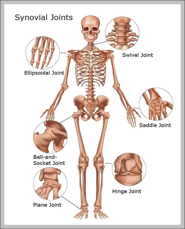
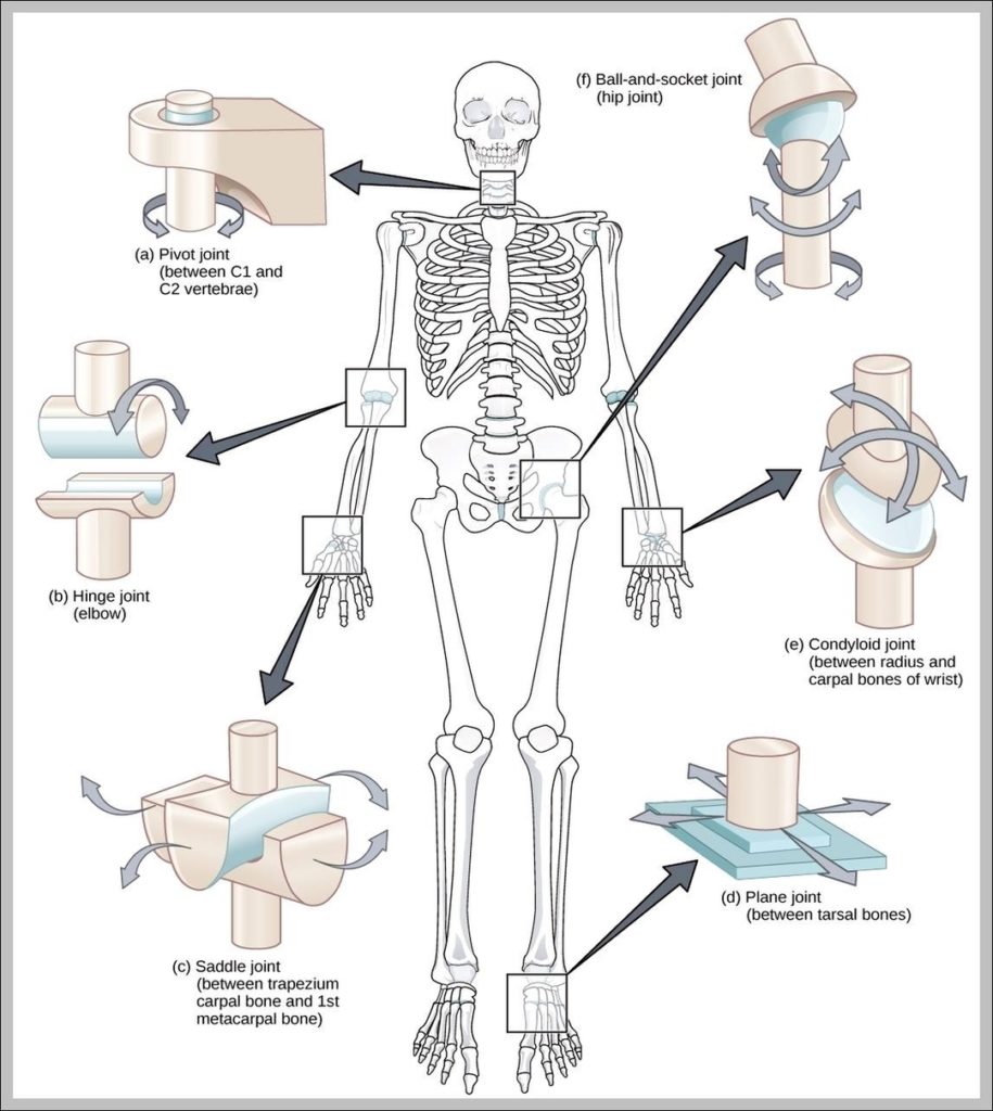
A hinge joint is a common class of synovial joint that includes the ankle, elbow, and knee joints. Hinge joints are formed between two or more bones where the bones can only move along one axis to flex or extend.
The elbows, the knees, and the middle and end joints of the fingers are hinge joints. Hinge joints are prone to injury when lateral forces are applied to the joint. Many knee injuries occur in this manner, according to the National Institute of Arthritis and Musculoskeletal and Skin Diseases.

Hinge Joints In The Body Image Diagram - Hinge Joints In The Body Image Chart - Human anatomy diagrams and charts explained. This anatomy system diagram depicts Hinge Joints In The Body Image with parts and labels. Best diagram to help learn about health, human body and medicine.

Hinge Joints In The Body Image

Joints In The Skeletal System Image

There are many types of tissues that make up joint anatomy. Typically, the bones that make up freely mobile joints are lined with articular (or hyaline) cartilage. Hyaline cartilage has a very smooth, almost frictionless surface, which allows for smooth, pain-free motion at the joint. Joints can be classified according to their degree of mobility:
Skeletal System: Bones, Joints, Cartilage, Ligaments, Bursae. The anatomy of our musculoskeletal system is quite complex. It consists of a large number of tendons, ligaments, bones, cartilage, joints, and bursae. We are able to control our muscles by sending stimulating impulses via nerves from our brain.

Joints In The Skeletal System Image Diagram - Joints In The Skeletal System Image Chart - Human anatomy diagrams and charts explained. This anatomy system diagram depicts Joints In The Skeletal System Image with parts and labels. Best diagram to help learn about health, human body and medicine.

Joints In The Skeletal System Image

Examples Of Hinge Joints Image

Examples Of Hinge Joints Image Diagram - Examples Of Hinge Joints Image Chart - Human anatomy diagrams and charts explained. This anatomy system diagram depicts Examples Of Hinge Joints Image with parts and labels. Best diagram to help learn about health, human body and medicine.

Examples Of Hinge Joints Image

Costovertebral Joints Anatomy Diagram

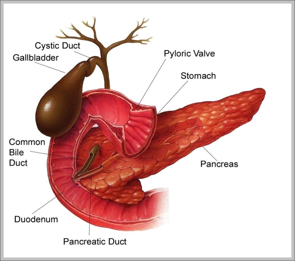
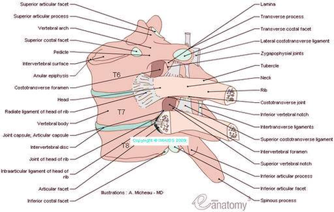
Costovertebral joint consists of the head of the rib (the head of a typical rib has two facets – each facet with a separate synovial joint separated by a ridge. The head of each rib articulates with: The lower rib facet articulates with the upper costal facet of its own vertebra
Costotransverse joint, seen from above. [edit on Wikidata] The costovertebral joints are the joints that connect the ribs to the vertebral column. The articulation of the head of the rib connects the head of the rib to the bodies of the thoracic vertebrae.
The connection between the heads of the ribs with the sides of one, or two adjacent vertebral bodies are known as the costocorporeal joints/joints of head of ribs. The articulations of the necks and tubercles of the ribs with the transverse processes of their corresponding thoracic vertebra are the costotransverse joints.

Costovertebral Joints Anatomy Diagram Diagram - Costovertebral Joints Anatomy Diagram Chart - Human anatomy diagrams and charts explained. This anatomy system diagram depicts Costovertebral Joints Anatomy Diagram with parts and labels. Best diagram to help learn about health, human body and medicine.

costovertebral joints anatomy diagram