Parts Of The Brain Diagram And Function

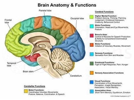
The human brain, the most complex organ in the body, is responsible for controlling thought, memory, emotion, touch, motor skills, vision, breathing, temperature, hunger, and every process that regulates our body. It is made up of billions of neurons and has several specialized parts, each involved in important functions.

1. Cerebrum
The cerebrum, the largest part of the brain, initiates and coordinates movement and regulates temperature. It comprises gray matter (the cerebral cortex) and white matter at its center. The cerebral cortex is responsible for higher-order functions such as consciousness, imagination, information processing, language, memory, perception, reasoning, sensation, and voluntary physical action. It is divided into four lobes:

– Frontal Lobe: Associated with reasoning, motor skills, higher-level cognition, and expressive language. Damage to this lobe can lead to changes in sexual habits, socialization, attention, and increased risk-taking.
– Parietal Lobe: Processes tactile sensory information such as pressure, touch, and pain.
– Occipital Lobe: Responsible for vision.
– Temporal Lobe: Interprets sounds and language we hear. It also heavily associated with the formation of memories.

2. Cerebellum
The cerebellum coordinates balance and muscle activity. It plays a vital role in motor control and is also involved in some cognitive functions such as attention and language, and in regulating fear and pleasure responses.

3. Brainstem
The brainstem controls basic functions like breathing. It acts as a relay center connecting the cerebrum and cerebellum to the spinal cord, performing many automatic functions such as breathing, heart rate, body temperature, wake and sleep cycles, digestion, sneezing, and coughing.

4. Diencephalon
The diencephalon regulates processes like sleep and body temperature. It consists of structures such as the thalamus and hypothalamus which are responsible for such functions as motor control, relaying sensory information, and controlling autonomic functions.

5. Limbic System
The limbic system, often referred to as the “emotional brain”, is involved in many of our emotions and motivations, particularly those related to survival such as fear and anger.

6. Gray and White Matter
Gray matter is primarily composed of neuron somas (the round central cell bodies), and white matter is mostly made of axons (the long stems that connect neurons together) wrapped in myelin (a protective coating). Gray matter is primarily responsible for processing and interpreting information, while white matter transmits that information to other parts of the nervous system.

In conclusion, the brain is a complex organ with various parts working together to regulate and control bodily functions. Each part plays a unique role, and understanding these parts can help us understand how disease and damage may affect the brain and its ability to function..

Parts Of The Brain Diagram And Function Diagram - Parts Of The Brain Diagram And Function Chart - Human anatomy diagrams and charts explained. This anatomy system diagram depicts Parts Of The Brain Diagram And Function with parts and labels. Best diagram to help learn about health, human body and medicine.
Parts Of The Brain Diagram And Function

Liver Location And Function Image

Here are a number of highest rated Location Of Major Organs Human Body pictures on internet. We identified it from well-behaved source. Its submitted by processing in the best field. We assume this kind of Location Of Major Organs Human Body graphic could possibly be the most trending topic considering we share it in google help or facebook.
80,662 human internal organ stock photos and images available, or search for human internal organ icons or human internal organ illustrations to find more great stock photos and pictures.

Liver Location And Function Image Diagram - Liver Location And Function Image Chart - Human anatomy diagrams and charts explained. This anatomy system diagram depicts Liver Location And Function Image with parts and labels. Best diagram to help learn about health, human body and medicine.
Liver Location And Function Image

Function Of The Lungs Image

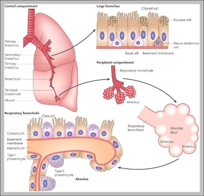
The lungs play an important part in the respiratory system. Their main function is to provide oxygen to the blood and remove carbon dioxide from the blood. Various diseases can affect the lungs, including asthma, infections, COPD, and cancer. Keep the lungs healthy by avoiding pollutants, getting regular exercise, and ceasing smoking.
Lung function tests, also known as pulmonary function tests, check how well the lungs work and can help investigate breathing problems. For example, tests can help screen and diagnose lung conditions such as asthma and COPD. They can also measure levels of function, monitor lung conditions, and see if treatments are working.

Function Of The Lungs Image Diagram - Function Of The Lungs Image Chart - Human anatomy diagrams and charts explained. This anatomy system diagram depicts Function Of The Lungs Image with parts and labels. Best diagram to help learn about health, human body and medicine.
Function Of The Lungs Image

Function Of Corpus Callosum Image

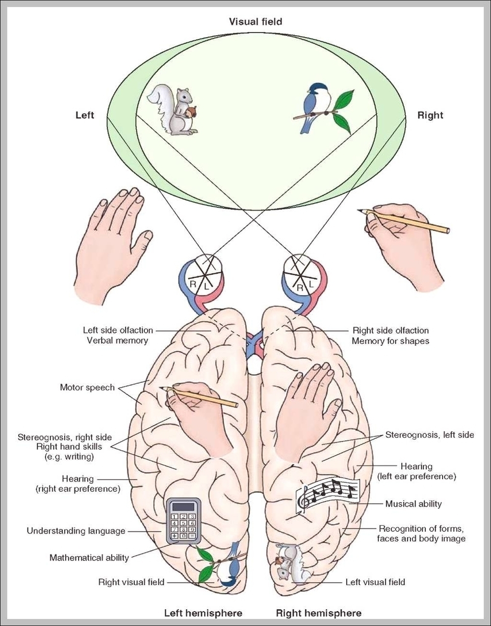
The corpus callosum is an important structure in the brain that plays a major role in nervous system communication, as well as contributing to its own important functions. In this lesson, investigate how the corpus callosum, its location, and its functions contribute to a well-functioning brain. Are you a student or a teacher?
Besides its importance in communication, the corpus callosum is hypothesized to play a primary role in cognition. Emerging evidence suggests that weakened integrity of the callosum contributes to a decline in cognitive function in aging adults.

Function Of Corpus Callosum Image Diagram - Function Of Corpus Callosum Image Chart - Human anatomy diagrams and charts explained. This anatomy system diagram depicts Function Of Corpus Callosum Image with parts and labels. Best diagram to help learn about health, human body and medicine.
Function Of Corpus Callosum Image

Function Of The Thyroid Gland Image

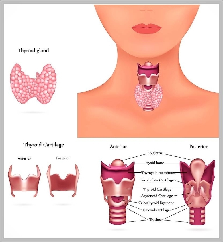
Thyroid gland. Medically reviewed by Healthline’s Medical Network on February 18, 2015. The thyroid gland covers the windpipe from three sides. Two hormones of the thyroid gland, T4 (thyroxine) and T3 (triiodothyronine), help the body to produce and regulate the hormones adrenaline (also called epinephrine) and dopamine.
The thyroid releases them into your bloodstream, which takes them through your body. Another gland, called the pituitary gland, tells your thyroid how much of these hormones your body needs. Sometimes, the thyroid gland develops a problem.

Function Of The Thyroid Gland Image Diagram - Function Of The Thyroid Gland Image Chart - Human anatomy diagrams and charts explained. This anatomy system diagram depicts Function Of The Thyroid Gland Image with parts and labels. Best diagram to help learn about health, human body and medicine.
Function Of The Thyroid Gland Image

Circulatory System Function Image

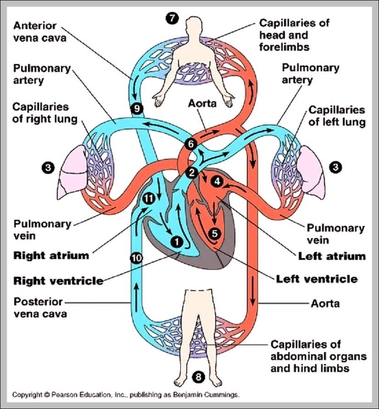
The circulatory system’s function is to move blood throughout the body. This blood circulation keeps organs, muscles and tissues healthy and working to keep you alive. The circulatory system also helps your body get rid of waste products. This waste includes: Carbon dioxide from respiration (breathing). Other chemical byproducts from your organs.
A fully functional circulatory system aims to maintain adequate concentration of oxygen in the biological tissues to ensure longevity and health. Circulatory system diagram can tell us clearly which parts are in play in the human circulation system. The circulatory system is primarily composed of:

Circulatory System Function Image Diagram - Circulatory System Function Image Chart - Human anatomy diagrams and charts explained. This anatomy system diagram depicts Circulatory System Function Image with parts and labels. Best diagram to help learn about health, human body and medicine.
Circulatory System Function Image

Function Of Capillaries Image

Function Of Capillaries Image Diagram - Function Of Capillaries Image Chart - Human anatomy diagrams and charts explained. This anatomy system diagram depicts Function Of Capillaries Image with parts and labels. Best diagram to help learn about health, human body and medicine.
Function Of Capillaries Image

Pulmonary Trunk Function Image

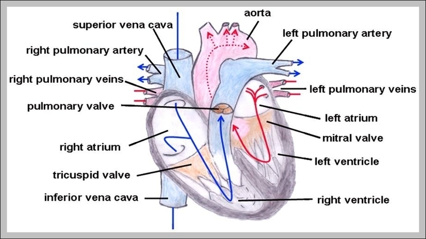
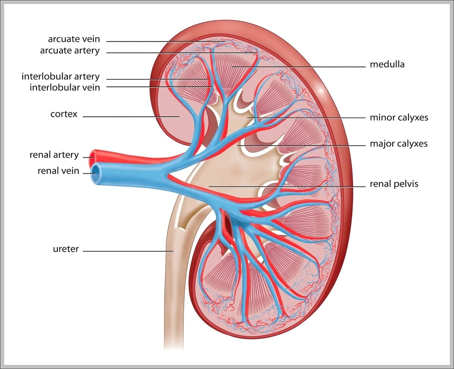
Pulmonary trunk. The pulmonary trunk or main pulmonary artery is the solitary arterial output from the right ventricle, transporting deoxygenated blood to the lungs for oxygenation.
1 Structure. The pulmonary trunk and the right and left pulmonary arteries are shaped somewhat like a capital letter “T”, with the trunk forming the lower portion and the left and … 2 Location. The pulmonary trunk, which is relatively short and wide, is located at the exit of the right ventricle. 3 Anatomic Variations. …

Pulmonary Trunk Function Image Diagram - Pulmonary Trunk Function Image Chart - Human anatomy diagrams and charts explained. This anatomy system diagram depicts Pulmonary Trunk Function Image with parts and labels. Best diagram to help learn about health, human body and medicine.
Pulmonary Trunk Function Image

Function Of The Kidney Image

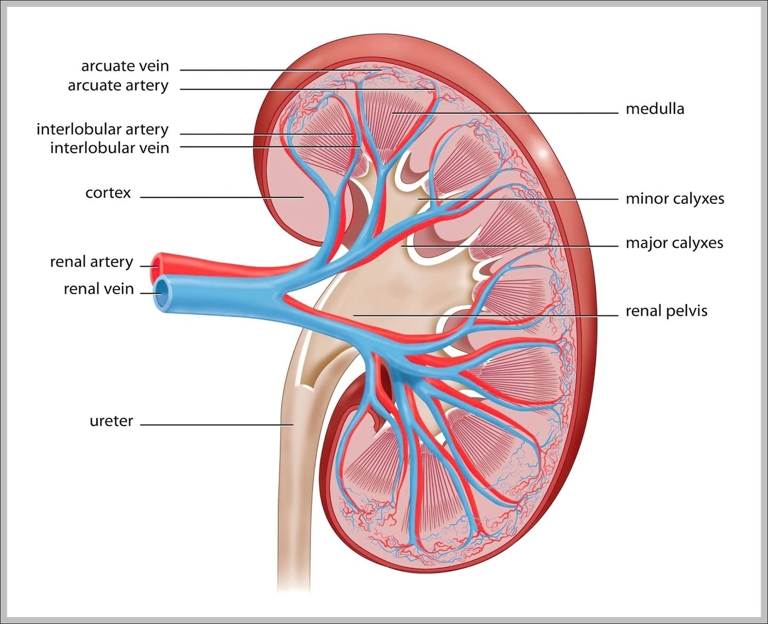
Most people know that the primary function of the kidneys is to eliminate waste products from the body by flushing them out with urine. However, did you know that there are at least 6 other fabulous functions you should thank your kidneys for?
A a Kidney Function Profile, is used to test the health of the kidneys. The nephron is the filtration unit of the kidney. It is the basic structural and functional unit of the kidney and cannot be seen by the naked eye. Each kidney has about 1 million nephrons. The walls of the nephron are made of a single layer of epithelial cells.

Function Of The Kidney Image Diagram - Function Of The Kidney Image Chart - Human anatomy diagrams and charts explained. This anatomy system diagram depicts Function Of The Kidney Image with parts and labels. Best diagram to help learn about health, human body and medicine.
Function Of The Kidney Image

Function Of Kidney Image

Most people know that the primary function of the kidneys is to eliminate waste products from the body by flushing them out with urine. However, did you know that there are at least 6 other fabulous functions you should thank your kidneys for?
A a Kidney Function Profile, is used to test the health of the kidneys. The nephron is the filtration unit of the kidney. It is the basic structural and functional unit of the kidney and cannot be seen by the naked eye. Each kidney has about 1 million nephrons. The walls of the nephron are made of a single layer of epithelial cells.

Function Of Kidney Image Diagram - Function Of Kidney Image Chart - Human anatomy diagrams and charts explained. This anatomy system diagram depicts Function Of Kidney Image with parts and labels. Best diagram to help learn about health, human body and medicine.

Function Of Kidney Image

Function Of The Pancreas Image

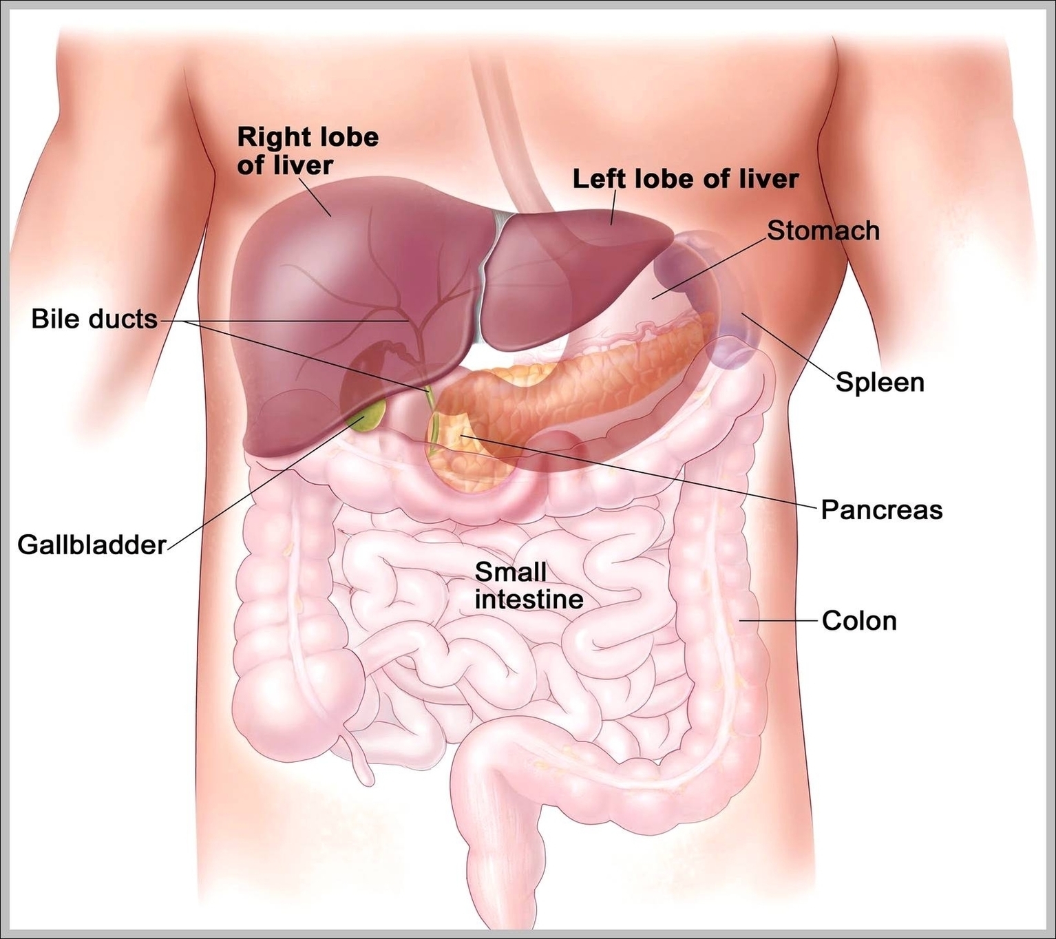
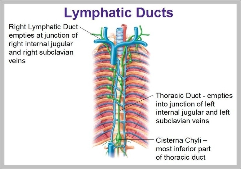
The pancreas is an organ located in the abdomen. It plays an essential role in converting the food we eat into fuel for the body’s cells. The pancreas has two main functions: an exocrine function that helps in digestion and an endocrine function that regulates blood sugar. The pancreas is located behind the stomach in the upper left abdomen.
Front View of the Pancreas. The head of the pancreas is on the right side of the abdomen and is connected to the duodenum (the first section of the small intestine) through a small tube called the pancreatic duct. The narrow end of the pancreas, called the tail, extends to the left side of the body.

Function Of The Pancreas Image Diagram - Function Of The Pancreas Image Chart - Human anatomy diagrams and charts explained. This anatomy system diagram depicts Function Of The Pancreas Image with parts and labels. Best diagram to help learn about health, human body and medicine.
Function Of The Pancreas Image

Thoracic Duct Function Image

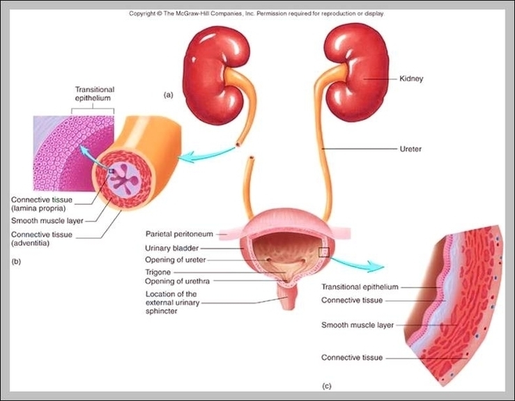
The thoracic duct carries chyle, a liquid containing both lymph and emulsified fats, rather than pure lymph. It also collects most of the lymph in the body other than from the right thorax, arm, head, and neck (which are drained by the right lymphatic duct ).
Variant anatomy. The thoracic duct has variant anatomy in ~40% (range 30-50%) of the population 1, 4: double thoracic ducts (i.e. multiple ducts) (~20%) 1, 2, 4 aberrant termination: left internal jugular vein, left external jugular vein, azygos vein, brachiocephalic vein or left subclavian vein 1, 4.

Thoracic Duct Function Image Diagram - Thoracic Duct Function Image Chart - Human anatomy diagrams and charts explained. This anatomy system diagram depicts Thoracic Duct Function Image with parts and labels. Best diagram to help learn about health, human body and medicine.
Thoracic Duct Function Image

Ureter Function Image

The ureter is a tube that carries urine from the kidney to the urinary bladder. There are two ureters, one attached to each kidney. … Duplication of the ureter: a congenital (from birth) condition in which two ureters form on the same kidney.
Gross anatomy. The ureter is 25-30 cm long and has three parts: abdominal ureter: from the renal pelvis to the pelvic brim.

Ureter Function Image Diagram - Ureter Function Image Chart - Human anatomy diagrams and charts explained. This anatomy system diagram depicts Ureter Function Image with parts and labels. Best diagram to help learn about health, human body and medicine.
Ureter Function Image

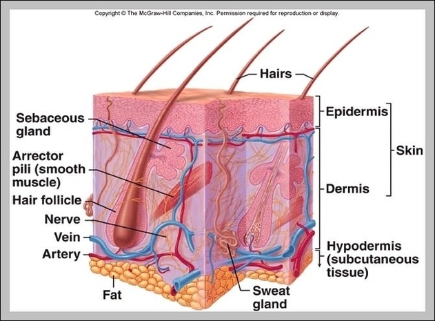
What Is The Function Of The Integumentary System Image

The primary function of the integumentary system is to protect the inside of the body from elements in the environment—like bacteria, pollution, and UV rays from the sun. The skin and its associated structures also retain bodily fluids, eliminate waste products, and regulate the body’s temperature.
The integumentary system helps regulate body temperature through its tight association with the sympathetic nervous system, the division of the nervous system involved in our fight-or-flight responses.

What Is The Function Of The Integumentary System Image Diagram - What Is The Function Of The Integumentary System Image Chart - Human anatomy diagrams and charts explained. This anatomy system diagram depicts What Is The Function Of The Integumentary System Image with parts and labels. Best diagram to help learn about health, human body and medicine.
What Is The Function Of The Integumentary System Image

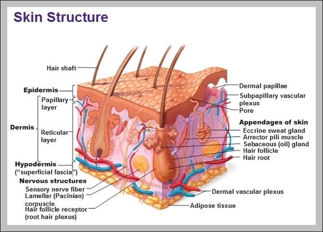
Function Of The Integumentary System Image

The primary function of the integumentary system is to protect the inside of the body from elements in the environment—like bacteria, pollution, and UV rays from the sun. The skin and its associated structures also retain bodily fluids, eliminate waste products, and regulate the body’s temperature.
The integumentary system helps regulate body temperature through its tight association with the sympathetic nervous system, the division of the nervous system involved in our fight-or-flight responses. The sympathetic nervous system is continuously monitoring body temperature and initiating appropriate motor responses.

Function Of The Integumentary System Image Diagram - Function Of The Integumentary System Image Chart - Human anatomy diagrams and charts explained. This anatomy system diagram depicts Function Of The Integumentary System Image with parts and labels. Best diagram to help learn about health, human body and medicine.

Function Of The Integumentary System Image

Function Of Veins Image

Vein Function. A vein is an elastic blood vessel that transports blood from various regions of the body to the heart. Veins can be categorized into four main types: pulmonary, systemic, superficial, and deep veins. Pulmonary veins carry oxygenated blood from the lungs to the heart. Systemic veins return deoxygenated blood from the rest…
Veins are thin vessels that are composed of three layers: Veins are different from arteries, which are muscular blood vessels that carry blood from the heart out to the body.

Function Of Veins Image Diagram - Function Of Veins Image Chart - Human anatomy diagrams and charts explained. This anatomy system diagram depicts Function Of Veins Image with parts and labels. Best diagram to help learn about health, human body and medicine.
Function Of Veins Image

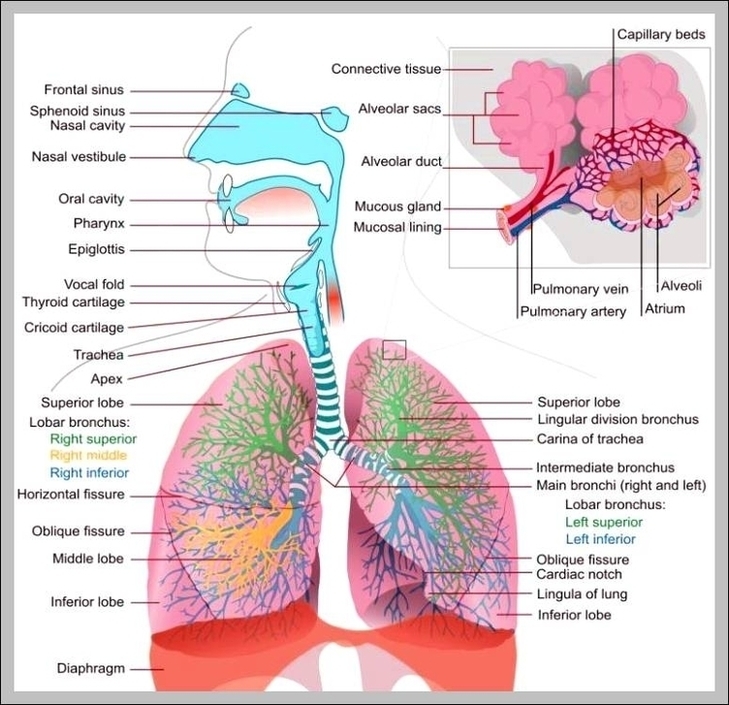
Function Of The Respiratory System Image

What are the Primary Functions of the Respiratory System Being one of the major biological systems, it serves a number of purposes in the human body, with three of its main functions being: Gas Exchange – A vital life-sustaining process where we inhale oxygen (O 2) and exhale carbon dioxide (CO 2) [1, 2].
Respiratory System icon. Trendy flat vector Respiratory System icon on white background from Human Body Parts collection, vector illustration can be use for web and mobile, eps10 Alveoli anatomy, oxygen and carbon dioxide exchange between alveoli and capillaries, external respiration mechanism.

Function Of The Respiratory System Image Diagram - Function Of The Respiratory System Image Chart - Human anatomy diagrams and charts explained. This anatomy system diagram depicts Function Of The Respiratory System Image with parts and labels. Best diagram to help learn about health, human body and medicine.

Function Of The Respiratory System Image

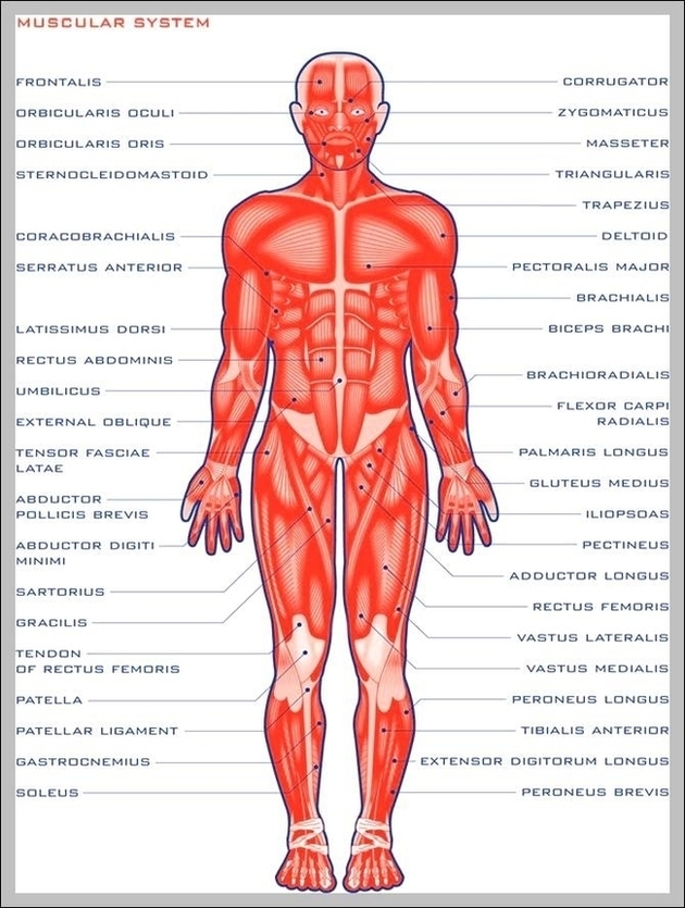
Deltoid Function Image

Function. The deltoid moves and stabilizes the shoulder joint. The movements of the different deltoid parts can interact both synergistically and antagonistically depending on the specific part and the position of the humerus. The deltoid is the most important abductor of the shoulder joint.
Anatomical terms of muscle. The deltoid muscle is the muscle forming the rounded contour of the human shoulder. It is also known as the common shoulder muscle, particularly in other animals such as the domestic cat.

Deltoid Function Image Diagram - Deltoid Function Image Chart - Human anatomy diagrams and charts explained. This anatomy system diagram depicts Deltoid Function Image with parts and labels. Best diagram to help learn about health, human body and medicine.
Deltoid Function Image

Major Function Of Muscular System Image

The main function of the muscular system is movement. Muscles are the only tissue in the body that has the ability to contract and therefore move the other parts of the body. Related to the function of movement is the muscular system’s second function: the maintenance of posture and body position.
The most obvious function of the muscular system is movement. Organisms have adopted a variety of methods to use the contractile function of the muscular system to move through the environment. The most basic movements of fish include contracting muscles on opposite sides of the body in succession.

Major Function Of Muscular System Image Diagram - Major Function Of Muscular System Image Chart - Human anatomy diagrams and charts explained. This anatomy system diagram depicts Major Function Of Muscular System Image with parts and labels. Best diagram to help learn about health, human body and medicine.
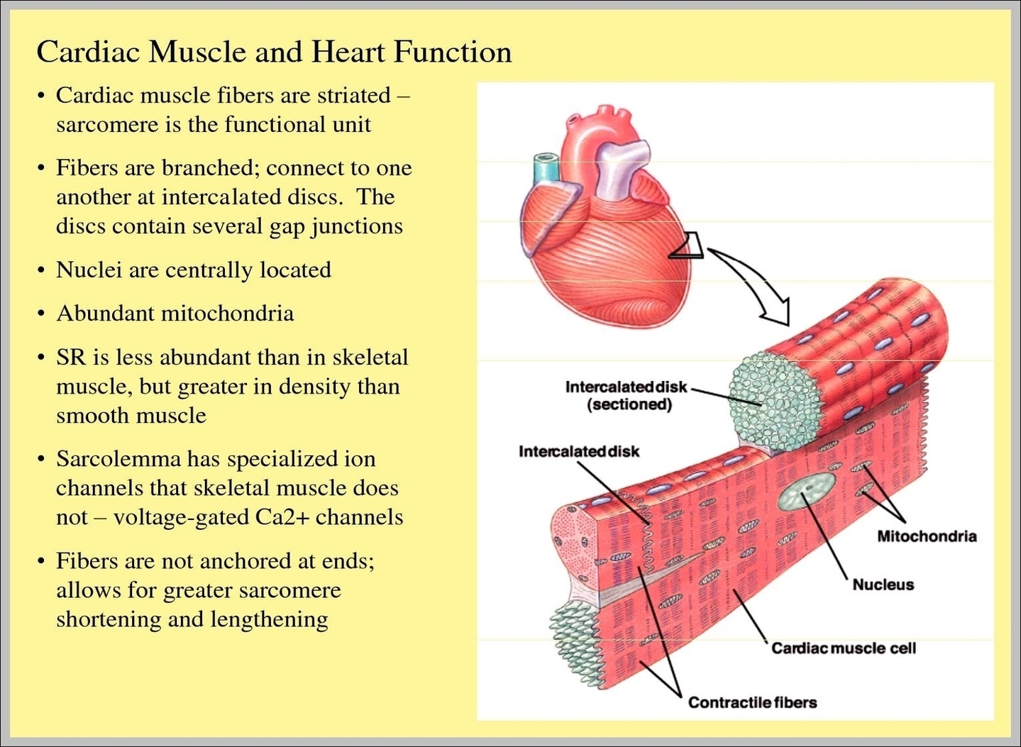
Major Function Of Muscular System Image

Cardiac Tissue Function Image

Cardiac muscle tissue is only found in your heart, where it performs coordinated contractions that allow your heart to pump blood through your circulatory system. Keep reading to learn more about the function and structure of cardiac muscle tissue, as well as conditions that affect this type of muscle tissue. How does it function?
Cardiac Muscle Definition Cardiac muscle, also known as heart muscle, is the layer of muscle tissue which lies between the endocardium and epicardium. These inner and outer layers of the heart, respectively, surround the cardiac muscle tissue and separate it from the blood and other organs.

Cardiac Tissue Function Image Diagram - Cardiac Tissue Function Image Chart - Human anatomy diagrams and charts explained. This anatomy system diagram depicts Cardiac Tissue Function Image with parts and labels. Best diagram to help learn about health, human body and medicine.

Cardiac Tissue Function Image