
Diagram Anatomy Ear
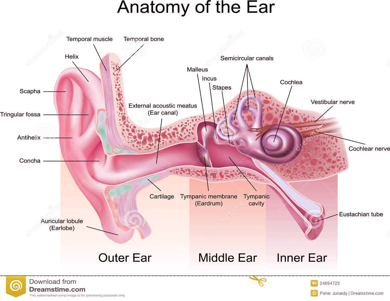
A brief description of the human ear along with a well-labelled diagram is given below for reference. Pinna/auricle is the outermost section of the ear. The external auditory canal links the exterior ear to the inner or the middle ear.
The middle ear is a chamber located within the petrous portion of the temporal bone. Structures within the middle ear amplify sound waves and transmit them to an appropriate portion of the internal ear. The internal ear contains the sensory organs for equilibrium (balance) and hearing. Figure 1. Ear structure Figure 2. Ear anatomy
All three parts of the ear are important for detecting sound by working together to move sound from the outer part through the middle and into the inner part of the ear. Ears also help to maintain balance. The outer part of the ear collects sound. Sound travels through the auricle and the auditory canal, a short tube that ends at the eardrum.