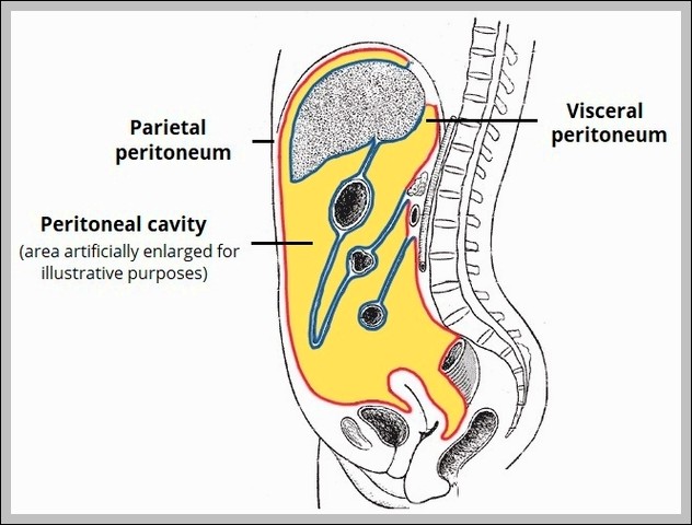
Overview of the Peritoneal Abdominal Cavity Diagram

Overview of the Peritoneal Abdominal Cavity Diagram

The peritoneal abdominal cavity includes parietal peritoneum lining walls, visceral covering organs, and mesenteries/omenta suspending structures. Greater/lesser sacs, transverse mesocolon, and root of mesentery divide spaces for fluid movement.
Overview of the Cerebrum1 Diagram

Overview of the Cerebrum Diagram

The cerebrum overview shows two hemispheres divided by longitudinal fissure, with frontal (planning, motor), parietal (sensory, spatial), temporal (auditory, memory), occipital (visual) lobes, plus insula deep in lateral sulcus and…
Overview of Parasympathetic Innervation Diagram

Overview of Parasympathetic Innervation Diagram

Parasympathetic innervation originates from cranial nerves (III, VII, IX, X) and sacral S2-S4. Preganglionic fibers synapse in terminal ganglia near targets for “rest and digest” functions: pupil constriction, salivation, lacrimation,…
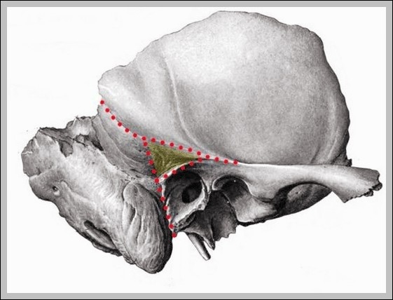
Mastoid Fossa Overview Diagram

Mastoid Fossa Overview Diagram

The mastoid fossa (antrum) is the largest mastoid air cell, opening into middle ear via aditus ad antrum, lined by mucosa continuous with tympanic cavity. It provides space for middle…
Overview Of The Digestive System

Overview Of The Digestive System

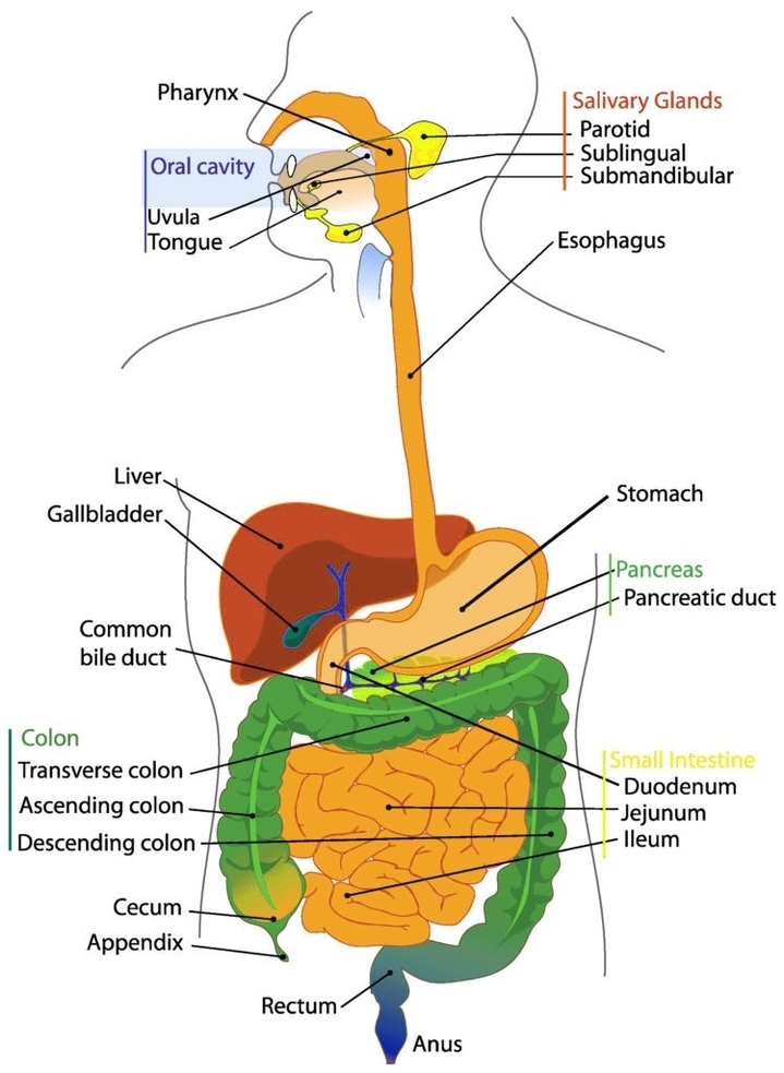
Overview of the Digestive System The digestive system is a complex network of organs that work together to break down food, absorb nutrients, and eliminate waste. It is composed of…