


Posted inMedical
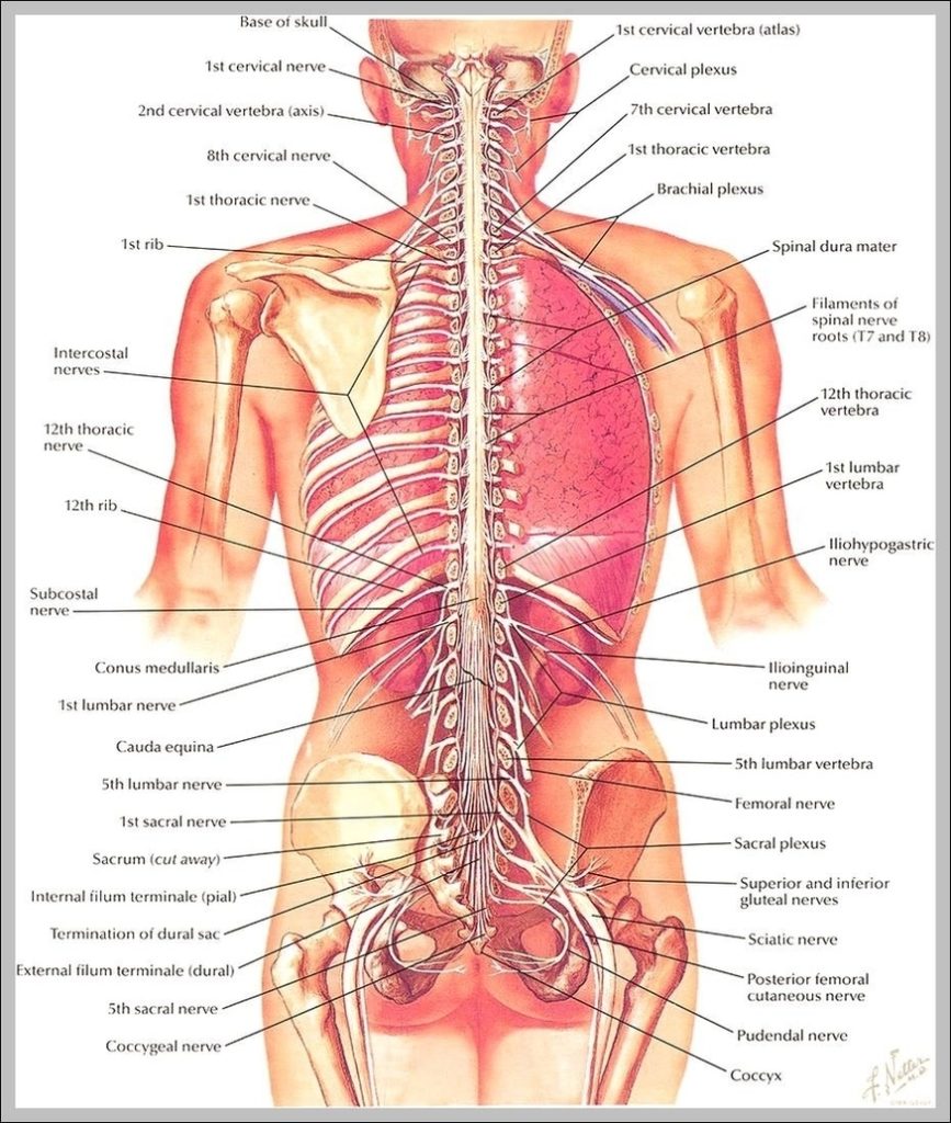
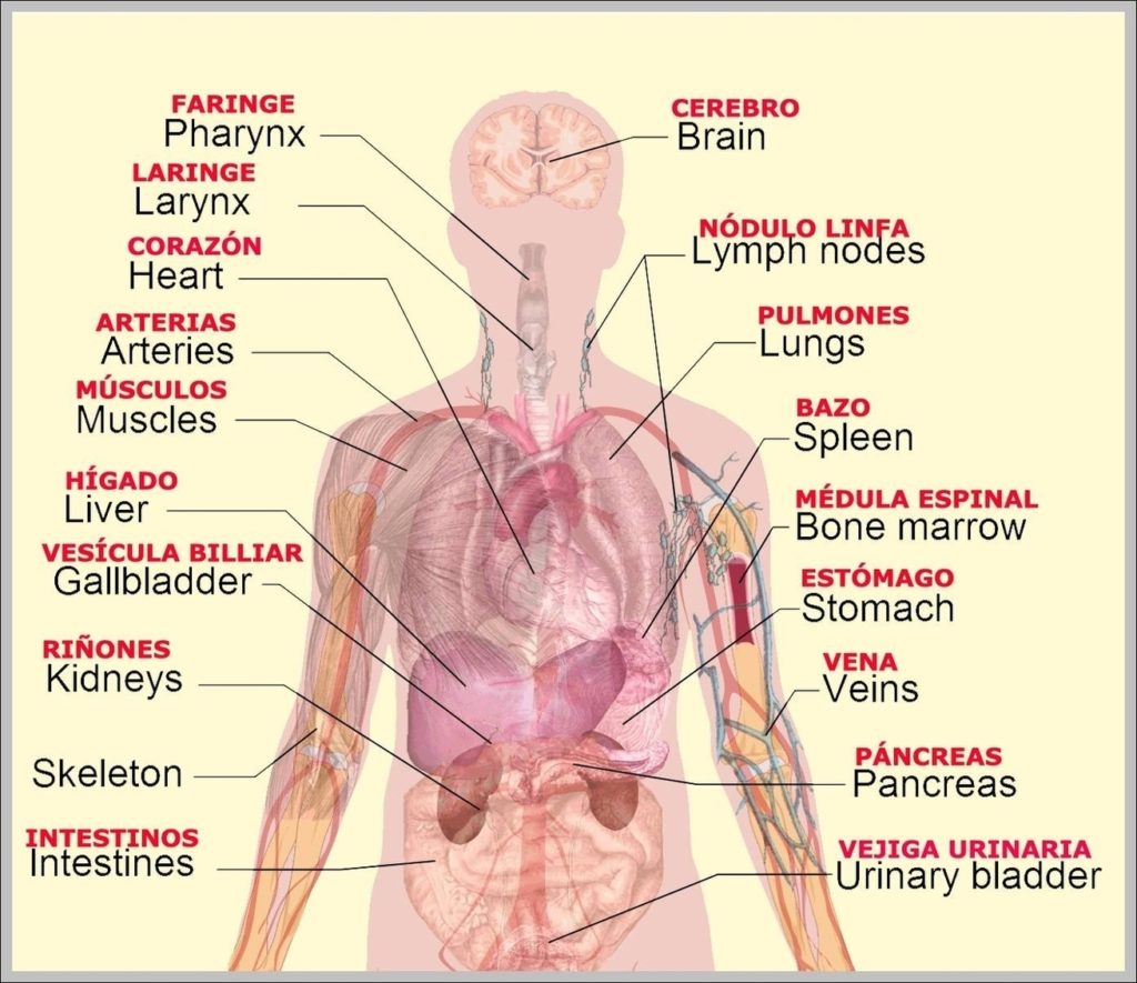
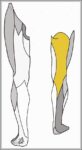
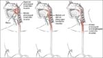
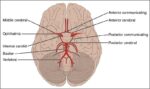
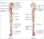
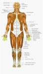
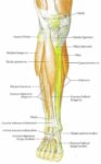
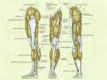
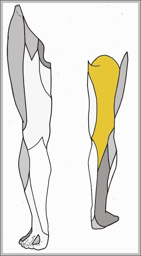
Posterior Cutaneous Nerve of Thigh Diagram
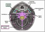
Posterior cutaneous nerve of thigh (S1-S3 posterior rami) supplies skin over posterior thigh and gluteal fold, running with sciatic nerve initially then superficially.