The Structure of a Vein Wall diagram layers out the typical three-part construction: a thin tunica intima with endothelium for smooth flow, a thicker tunica media with some smooth muscle…
The long thoracic nerve (C5-C7) descends superficially on serratus anterior, supplying it for scapular protraction/rotation. Injury causes winged scapula and difficulty raising arm overhead.
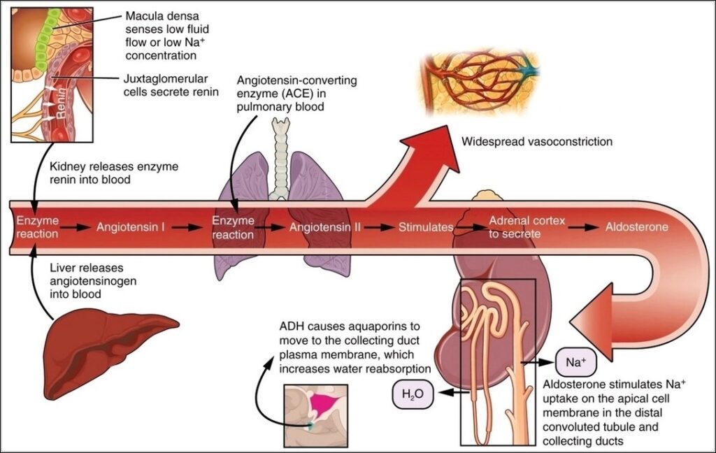
Renin-angiotensin-aldosterone system activates on low renal perfusion sensed by juxtaglomerular cells releasing renin converting angiotensinogen to angiotensin I, ACE in lungs to II potent vasoconstrictor raising pressure, stimulating aldosterone from…
Parasympathetic innervation originates from cranial nerves (III, VII, IX, X) and sacral S2-S4. Preganglionic fibers synapse in terminal ganglia near targets for rest and digest functions: pupil constriction, salivation, lacrimation,…
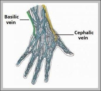
Superficial upper limb veins originate as dorsal venous arch (cephalic lateral to basilic medial), draining to median cubital at elbow, then axillary/subclavian. Theyre key for IV access.
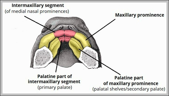
The development of the palate begins with primary palate formation from medial nasal prominences (intermaxillary segment) anteriorly, followed by secondary palate from lateral palatal shelves of maxillary prominences. Shelves grow…
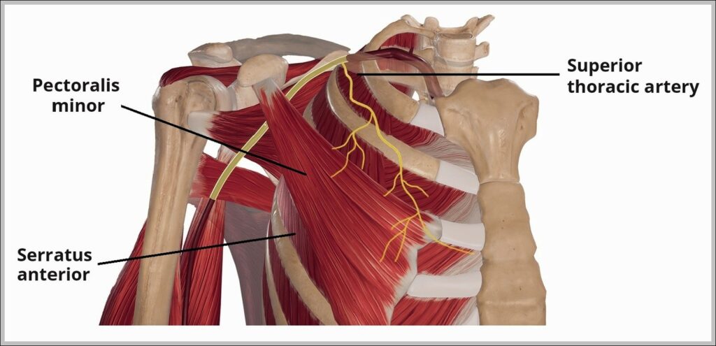
This Course of the Superior Thoracic Artery diagram traces this small branch from the first part of the subclavian artery, descending to supply the upper intercostal spaces, chest wall muscles,…
The posterior interosseous artery arises from common interosseous (from ulnar), runs with posterior interosseous nerve through supinator, supplies posterior forearm extensors. The supinator muscle wraps radius, innervated by deep radial…
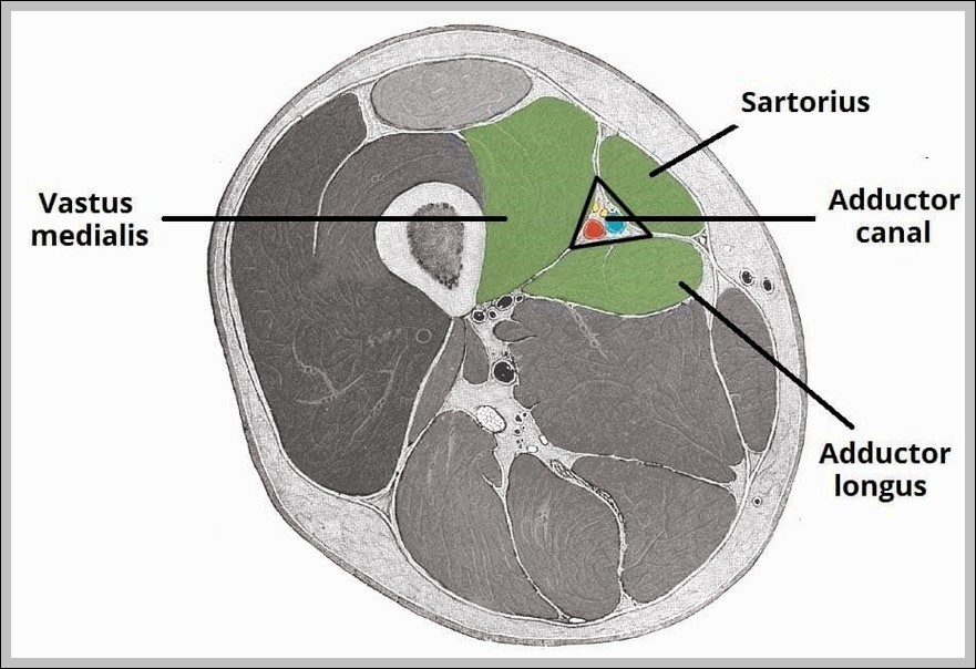
The Borders and Contents of the Adductor Canal diagram defines this subsartorial tunnel from apex of femoral triangle to adductor hiatus: roof (sartorius), anterolateral (vastus medialis), posteromedial (adductors longus/magnus). Contents…
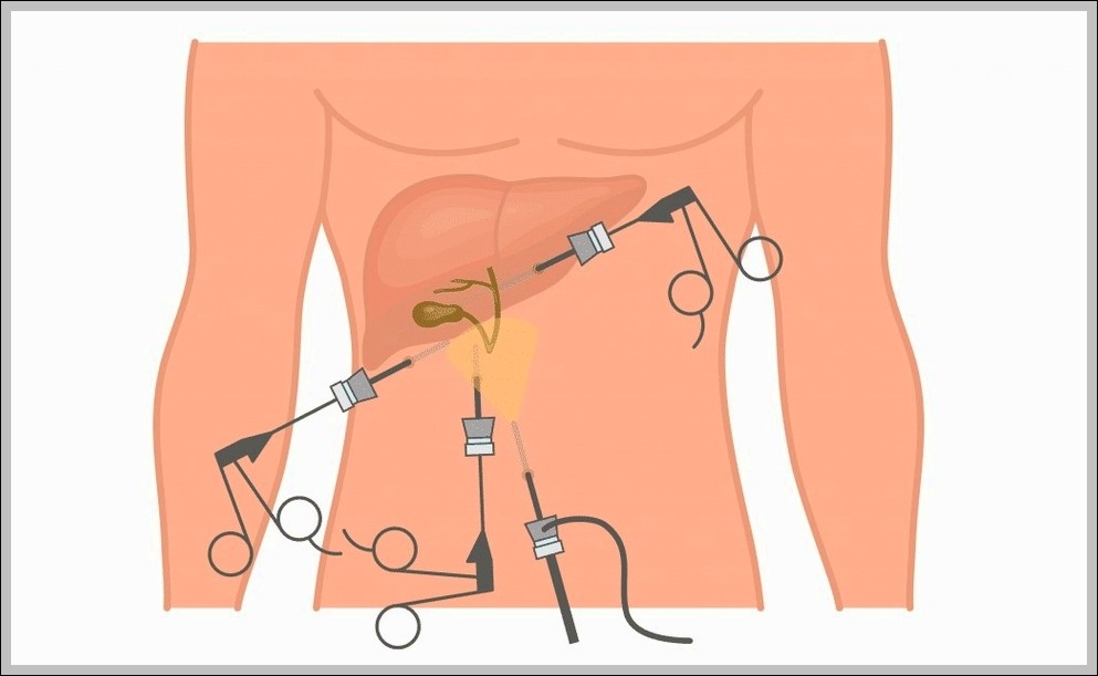
This Laparoscopic Cholecystectomy diagram shows the key steps of gallbladder removal using small incisions and a camera. It highlights where the trocars go, the tools in action, and important structures…
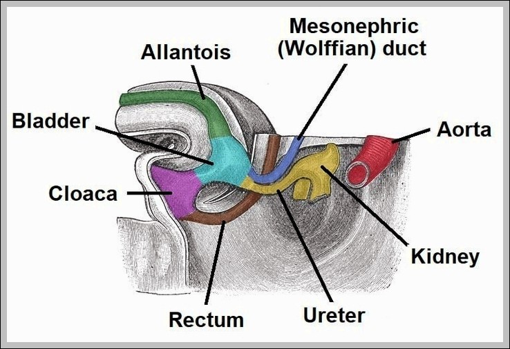
The Development of the Bladder Ureter Mesonephric Duct diagram traces embryonic stages where the mesonephric (Wolffian) duct gives rise to the ureteric bud, which induces metanephric blastema for kidney formation…
Neurulation forms the CNS from neural plate (ectoderm thickening) folding into neural groove, then neural tube by fusion of neural folds. Anterior neuropore closes first (day 25), posterior last (day…
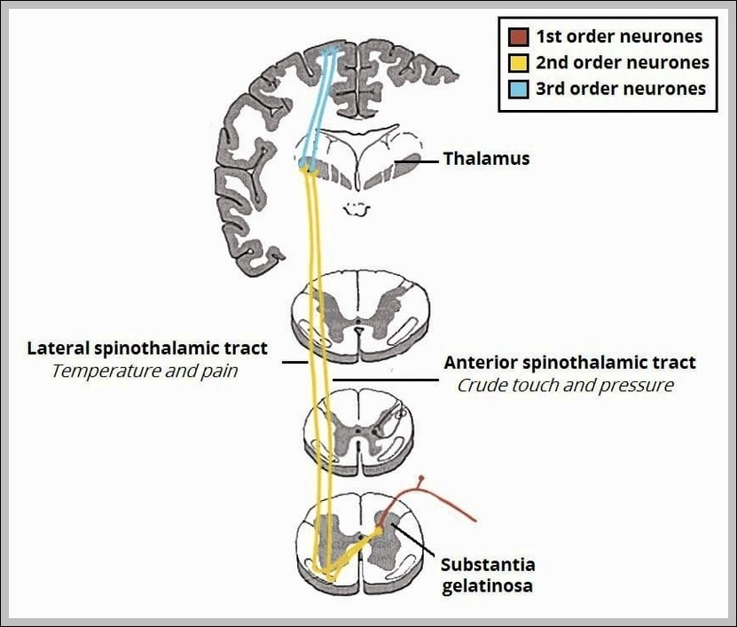
The spinothalamic tracts carry pain, temperature, and crude touch from the body: lateral for pain/temp, anterior for touch/pressure. Fibers cross in the spinal cord shortly after entry, ascend contralaterally to…
Nerves of dorsum foot include superficial fibular (sensory to dorsum except first webspace), deep fibular (first webspace skin, extensor digitorum brevis), sural (lateral foot). They emerge from anterior/lateral leg compartments.
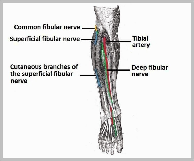
The common fibular (peroneal) nerve wraps around fibular neck (vulnerable to injury), divides into superficial (lateral compartment, peroneus longus/brevis) and deep (anterior compartment, tibialis anterior, extensors) branches. It supplies dorsiflexion,…