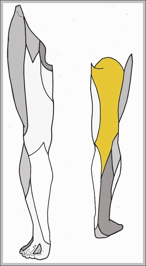
Histology of the Large IntestineN

Histology of the Large IntestineN

The large intestine's histology is geared toward water absorption, electrolyte balance, and waste formation, with a mucosa lacking villi but featuring deep crypts of Lieberkühn lined by columnar absorptive cells…
Fetal Circulatory System

Fetal Circulatory System

The fetal circulatory system is beautifully adapted to bypass the lungs and liver while maximizing placental exchange. Blood leaves the fetus through two umbilical arteries branching from the iliac arteries,…
Structures of the Respiratory Zone

Structures of the Respiratory Zone

Structures of respiratory zone begin distal terminal bronchioles with occasional alveoli respiratory bronchioles more leading alveolar ducts fully walled sacs clusters sharing openings millions thin-walled pouches type I cells diffusion…
Major Systemic Artery

Major Systemic Artery

Major systemic arteries begin with ascending aorta giving coronaries, arch branching brachiocephalic, left common carotid, left subclavian, then descending thoracic supplying intercostals, abdominal continuing as celiac, mesenteric, renal, gonadal, iliacs,…
Implantation

Implantation

Implantation occurs about a week after fertilization when the blastocyst, having hatched from zona pellucida, adheres to endometrial lining primed by progesterone, with trophoblast cells invading to establish placental connections.…
Varicose Vein

Varicose Vein

Varicose veins are dilated tortuous superficial veins usually lower limbs from valve incompetence allowing reflux pooling under gravity, great saphenous common, risk factors standing pregnancy obesity heredity, symptoms aching heaviness…
Lower Limb Arteries Chart

Lower Limb Arteries Chart

The arterial supply to lower limbs forms a continuous chain with branches providing redundancy, starting with common femoral dividing into superficial femoral through adductor canal and profunda femoris laterally, then…
Heart Musculature

Heart Musculature

The heart's muscular wall, the myocardium, consists of specialized cardiac muscle cells arranged in spiral and circular bundles unique to each chamber. Atrial muscle is thinner and arranged in loops,…
Comparison of Artery and Vein

Comparison of Artery and Vein

A side-by-side vascular comparison highlights the structural differences between arteries and veins. Thick muscular walls, narrow lumens, and elastic layers are shown in arteries, while veins appear thinner with wider…
Stethoscope Placement

Stethoscope Placement

Stethoscope placement for heart sounds uses four standard sites: aortic second right intercostal sternal border high-pitched semilunar closures, pulmonic second left for same, tricuspid lower left sternal for right AV,…
Cooperation Between Innate and Immune Responses

Cooperation Between Innate and Immune Responses

An immune system interaction diagram illustrates how innate and adaptive responses work together. Early defenses such as macrophages and inflammation are shown activating and guiding lymphocytes. This cooperation explains faster…
Blood Flow Contracted Ventricles

Blood Flow Contracted Ventricles

A heart cycle image showing contracted ventricles focuses on blood being forced into the aorta and pulmonary artery. Valve positions and flow direction clarify how each heartbeat moves blood efficiently…
Gross Anatomy of the Lungs

Gross Anatomy of the Lungs

The lungs are paired spongy organs in the thoracic cavity, each divided into lobes—three on the right, two on the left—to accommodate the heart's position. The right lung is shorter…
Mucosa Associated Lymphoid Tissue MALT Nodule

Mucosa Associated Lymphoid Tissue MALT Nodule

Mucosa-associated lymphoid tissue, including nodules like Peyer’s patches in ileum, tonsils, and appendix, forms diffuse surveillance networks beneath epithelial surfaces rich in antigens, with M cells sampling lumen contents for…
Embroyonic Disc Amniotic Cavity Yolk Sac

Embroyonic Disc Amniotic Cavity Yolk Sac

The embryonic disc with amniotic cavity and yolk sac represents the early post-implantation stage, where the bilaminar disc of epiblast and hypoblast forms, then gastrulation creates a trilaminar disc with…
Cardiac Cycle vs Heart Sounds

Cardiac Cycle vs Heart Sounds

A comparison of the cardiac cycle and heart sounds visually links mechanical events in the heart to what is heard through a stethoscope. The diagram aligns phases such as atrial…
Internal Anatomy of the HeartN

Internal Anatomy of the Heart

The heart's internal anatomy features four chambers with septa dividing left and right, atria receiving blood via smooth posterior walls and pectinate anterior plus auricles, ventricles with trabeculae carneae and…
Major Muscles Of The Human Body

Major Muscles Of The Human Body

Major Muscles of the Human Body The human body is a complex system that comprises more than 600 muscles. These muscles are broadly divided into three types: 1. Skeletal Muscle:…