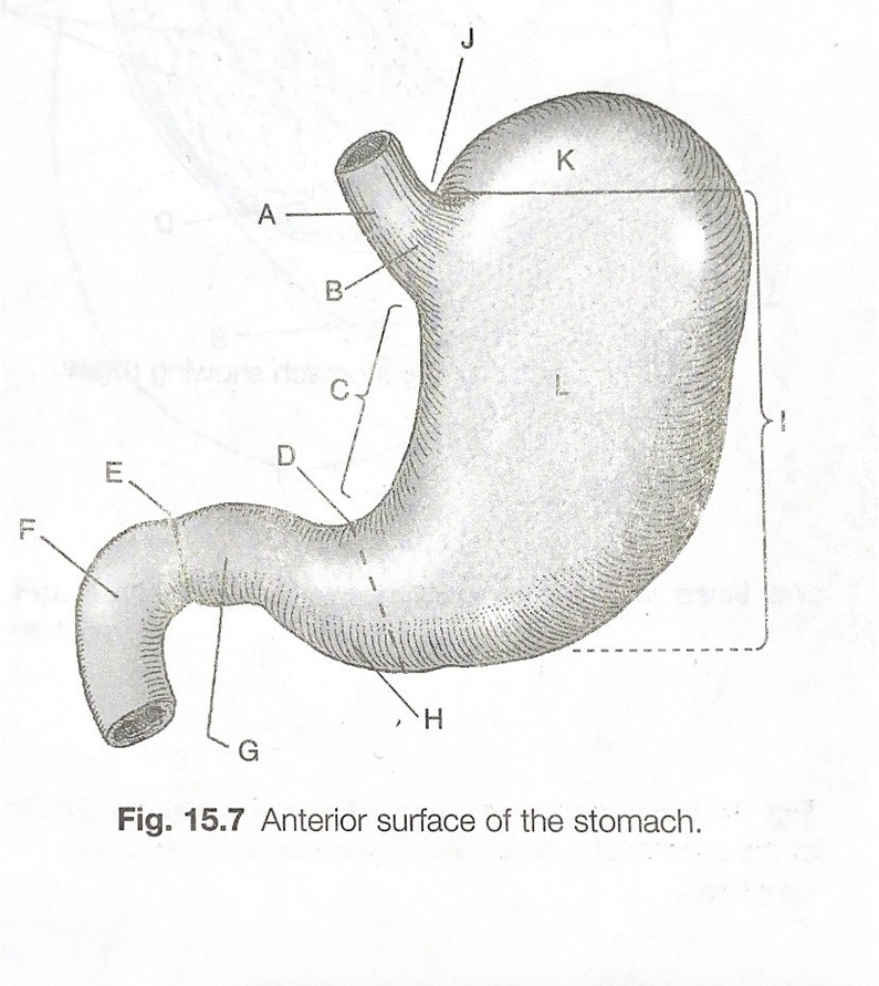
Stomach Diagram Graphic
The stomach is a muscular, hollow organ in the gastrointestinal tract of humans and many other animals. It is located in the upper left abdominal area and is part of the digestive system. The stomach serves as a temporary receptacle for the storage and mechanical distribution of food before it is passed into the intestine.
The stomach is a J-shaped organ that digests food. It produces enzymes (substances that create chemical reactions) and acids (digestive juices). This mix of enzymes and digestive juices breaks down food so it can pass to your small intestine. The stomach is part of the gastrointestinal (GI) tract. The GI tract is a long tube that starts at your mouth. It runs to your anus, where stool (poop) leaves your body. The GI tract is a key part of your digestive system.
The stomach’s purpose is to digest food and send it to your small intestine. It has three functions:
1. Temporarily store food.
2. Contract and relax to mix and break down food.
3. Produce enzymes and other specialized cells to digest food.
Each part of your GI tract breaks down food and liquid and carries it through your body. During the digestive process, your body absorbs nutrients and water. Then, you expel the waste products of digestion through your large intestine.
Food moves through your GI tract in a few steps:
1. Mouth: As you chew and swallow, your tongue pushes food into your throat. A small piece of tissue called the epiglottis covers your windpipe. The epiglottis prevents choking.
2. Esophagus: Food travels down a hollow tube called the esophagus. At the bottom, your esophageal sphincter relaxes to let food pass to your stomach.
3. Stomach: Your stomach creates digestive juices and breaks down food. It holds food until it is ready to empty into your small intestine.
4. Small intestine: Food mixes with the digestive juices from your intestine, liver, and pancreas. Your intestinal walls absorb nutrients and water from food and send waste products to the large intestine.
5. Large intestine: Your large intestine turns waste products into stool. It pushes the stool into your rectum.
6. Rectum: The rectum is the lower portion of your large intestine. It stores stool until you have a bowel movement.
The stomach is surrounded by parasympathetic (stimulant) and sympathetic (inhibitor) plexuses (networks of blood vessels and nerves in the anterior gastric, posterior, superior and inferior, celiac and myenteric), which regulate both the secretory activity of the stomach and the motor (motion) activity of its muscles.
Because it is a distensible organ, it normally expands to hold about one litre of food. The stomach of a newborn human baby will only be able to retain about 30 millilitres. The maximum stomach volume in adults is between 2 and 4 litres. Although volumes of up to 15 L have been observed in extreme circumstances.
In classical anatomy, the human stomach is divided into four sections, beginning at the cardia. The stomach secretes digestive enzymes and gastric acid to aid in food digestion. The pyloric sphincter controls the passage of partially digested food (chyme) from the stomach into the duodenum, where peristalsis takes over to move this through the rest of the intestines..
Stomach Diagram Graphic Diagram - Stomach Diagram Graphic Chart - Human anatomy diagrams and charts explained. This anatomy system diagram depicts Stomach Diagram Graphic with parts and labels. Best diagram to help learn about health, human body and medicine.