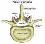


Posted inDiagrams
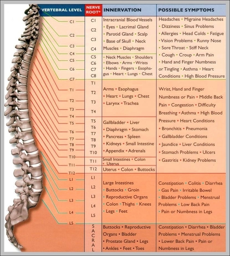
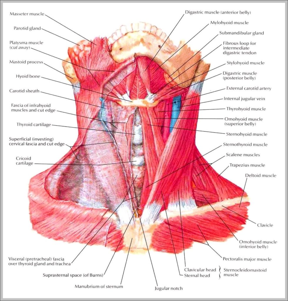
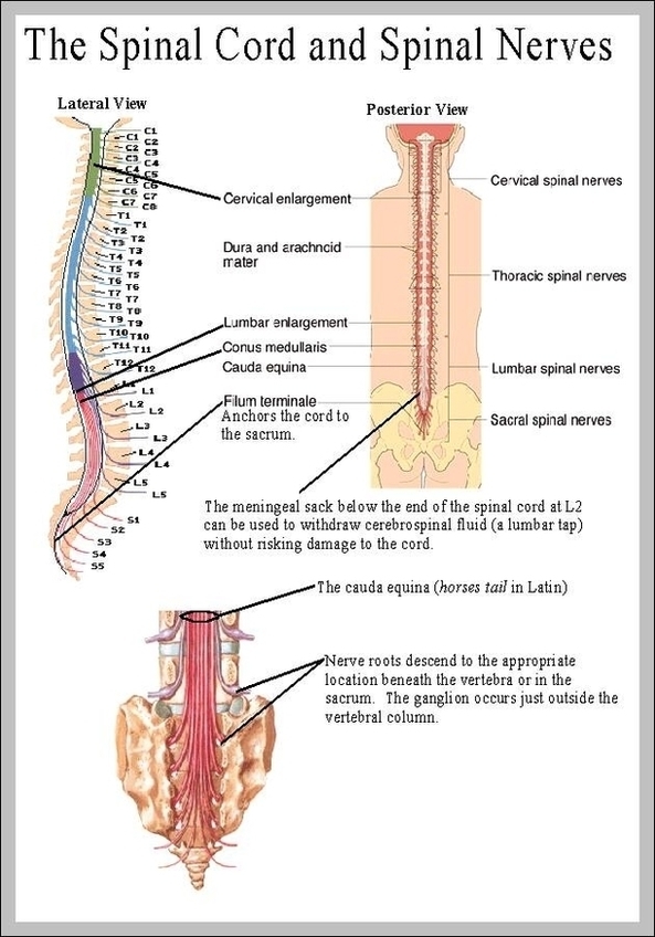
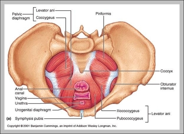
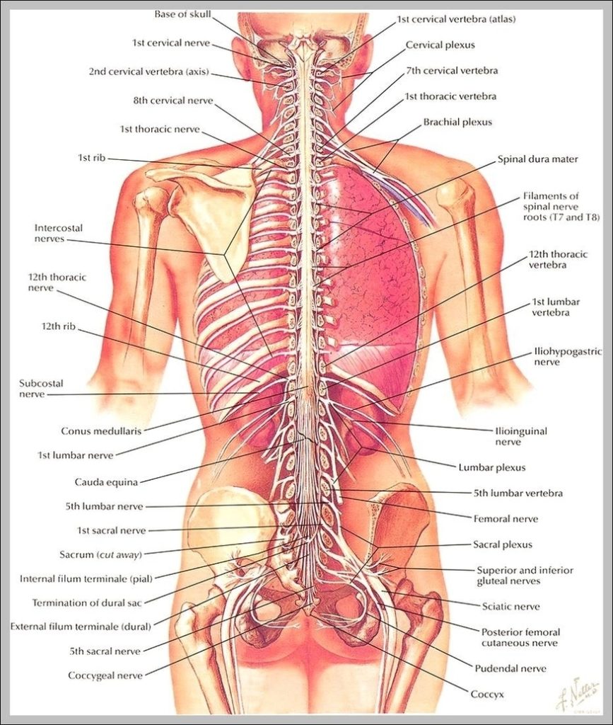
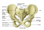
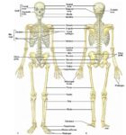
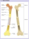
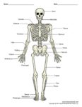
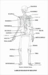
Human Vertebral Column Anatomy
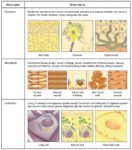
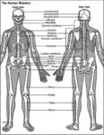
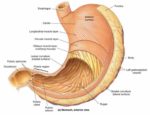
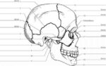
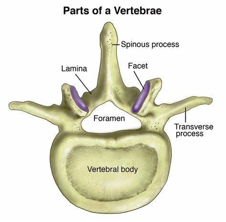
Human Vertebral Column Anatomy The human vertebral column, also known as the spine or backbone, is a crucial component of the human skeletal system. It is a curved structure composed…