Ear Anatomysimple Ear Anatomy Visual

Anatomy of the Human Ear

The human ear is a complex organ that serves two primary functions: hearing and maintaining balance. It is anatomically divided into three main parts: the outer ear, the middle ear, and the inner ear.

1. Outer Ear

The outer ear consists of the visible portion called the auricle, or pinna, and the external auditory canal. The auricle collects sound waves and guides them into the auditory canal. The inner end of the canal is closed by the tympanic membrane, commonly known as the eardrum. The function of the outer ear is to collect sound waves and guide them to the tympanic membrane.

2. Middle Ear

The middle ear is an air-filled cavity in the temporal bone. It contains a chain of three tiny bones — the malleus (hammer), incus (anvil), and stapes (stirrup), collectively known as the auditory ossicles. These bones conduct sound from the tympanic membrane to the inner ear.

3. Inner Ear

The inner ear, also known as the labyrinth, is a complex system of fluid-filled passages and cavities. It consists of two functional units: the vestibular apparatus and the cochlea. The vestibular apparatus, which includes the vestibule and semicircular canals, maintains balance. The cochlea, on the other hand, is responsible for hearing. These sensory organs are highly specialized endings of the eighth cranial nerve, also known as the vestibulocochlear nerve

Ear Anatomysimple Ear Anatomy Visual Diagram - Ear Anatomysimple Ear Anatomy Visual Chart - Human anatomy diagrams and charts explained. This anatomy system diagram depicts Ear Anatomysimple Ear Anatomy Visual with parts and labels. Best diagram to help learn about health, human body and medicine.

Ear Anatomysimple Ear Anatomy Visual

Brain Anatomy Diagram

Brain Anatomy

The brain, a complex organ, controls thought, memory, emotion, touch, motor skills, vision, breathing, temperature, hunger, and every process that regulates our body. It is primarily composed of nerve cells, also known as neurons. Blood vessels supply oxygen and nutrients to the neurons of the brain.

Composition

Weighing about 3 pounds in the average adult, the brain is about 60% fat. The remaining 40% is a combination of water, protein, carbohydrates, and salts. The brain itself is not a muscle. It contains blood vessels and nerves, including neurons and glial cells.

Gray Matter and White Matter

Gray and white matter are two different regions of the central nervous system. In the brain, gray matter refers to the darker, outer portion, while white matter describes the lighter, inner section underneath. Gray matter is primarily composed of neuron somas (the round central cell bodies), and white matter is mostly made of axons (the long stems that connect neurons together) wrapped in myelin (a protective coating). Gray matter is primarily responsible for processing and interpreting information, while white matter transmits that information to other parts of the nervous system.

Brain Function

The brain sends and receives chemical and electrical signals throughout the body. Different signals control different processes, and your brain interprets each. Some make you feel tired, for example, while others make you feel pain. Some messages are kept within the brain, while others are relayed through the spine and across the body’s vast network of nerves to distant extremities.

Main Parts of the Brain and Their Functions

At a high level, the brain can be divided into the cerebrum, brainstem, and cerebellum.

1. Cerebrum: The cerebrum (front of brain) comprises gray matter (the cerebral cortex) and white matter at its center. The largest part of the brain, the cerebrum initiates and coordinates movement and regulates temperature. Other areas of the cerebrum enable speech, judgment, thinking and reasoning, problem-solving, emotions, and learning. Other functions relate to vision, hearing, touch, and other senses.

2. Cerebral Cortex: The cortex has a large surface area due to its folds, and comprises about half of the brain’s weight. The cerebral cortex is divided into two halves, or hemispheres. It is covered with ridges (gyri) and folds (sulci).

3. Brain Stem: The brain stem controls the flow of messages between the brain and the rest of the body, and it also controls basic body functions such as breathing, swallowing, heart rate, blood pressure, consciousness, and whether one is awake or sleepy.

4. Cerebellum: The cerebellum is involved in the coordination of voluntary motor movement, balance and equilibrium and muscle tone.

5. Limbic System: The limbic system is a complex set of structures that lies on both sides of the thalamus, just under the cerebrum. It includes the hypothalamus, the hippocampus, the amygdala, and several other nearby areas. It appears to be primarily responsible for our emotional life, and has a lot to do with the formation of memories.

In conclusion, the brain is a complex organ with various parts working together to regulate our body’s functions and processes. Understanding its anatomy helps us appreciate its importance and the role it plays in our daily lives..

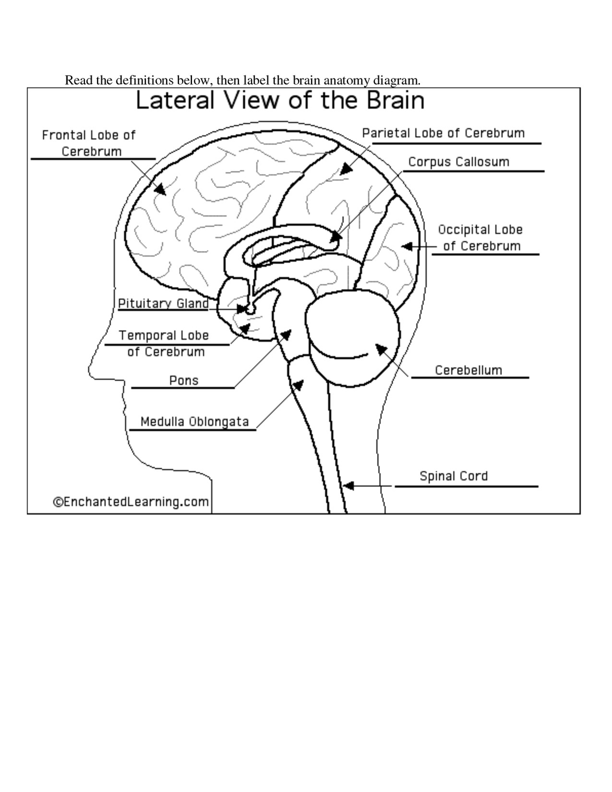
Brain Anatomy Diagram Diagram - Brain Anatomy Diagram Chart - Human anatomy diagrams and charts explained. This anatomy system diagram depicts Brain Anatomy Diagram with parts and labels. Best diagram to help learn about health, human body and medicine.

Brain Anatomy Diagram

Human Brain Anatomy Graphic

Human Brain Anatomy

The human brain, a complex organ, is the central component of the nervous system. It controls thought, memory, emotion, touch, motor skills, vision, breathing, temperature, hunger, and every process that regulates our body.

Composition

Weighing about 3 pounds in the average adult, the brain is about 60% fat. The remaining 40% is a combination of water, protein, carbohydrates, and salts. The brain is primarily composed of nerve cells, also known as neurons, and supportive glial cells. It contains blood vessels and nerves, including neurons and glial cells.

Gray Matter and White Matter

Gray and white matter are two different regions of the central nervous system. In the brain, gray matter refers to the darker, outer portion, while white matter describes the lighter, inner section underneath. Gray matter is primarily composed of neuron somas (the round central cell bodies), and white matter is mostly made of axons (the long stems that connect neurons together) wrapped in myelin (a protective coating). Gray matter is primarily responsible for processing and interpreting information, while white matter transmits that information to other parts of the nervous system.

Main Parts of the Brain

The brain can be divided into the cerebrum, brainstem, and cerebellum.

1. Cerebrum: The cerebrum (front of the brain) comprises gray matter (the cerebral cortex) and white matter at its center. The largest part of the brain, the cerebrum initiates and coordinates movement and regulates temperature. Other areas of the cerebrum enable speech, judgment, thinking and reasoning, problem-solving, emotions, and learning. Other functions relate to vision, hearing, touch, and other senses.

2. Cerebral Cortex: The cortex has a large surface area due to its folds, and comprises about half of the brain’s weight. The cerebral cortex is divided into two halves, or hemispheres. It is covered with ridges (gyri) and folds (sulci).

3. Brainstem: The brainstem consists of the midbrain, the pons, and the medulla oblongata.

4. Cerebellum: The cerebellum is connected to the brainstem by three pairs of nerve tracts called cerebellar peduncles.

Functioning

The brain sends and receives chemical and electrical signals throughout the body. Different signals control different processes, and your brain interprets each. Some make you feel tired, for example, while others make you feel pain. Some messages are kept within the brain, while others are relayed through the spine and across the body’s vast network of nerves to distant extremities. To do this, the central nervous system relies on billions of neurons (nerve cells)..

Human Brain Anatomy Graphic Diagram - Human Brain Anatomy Graphic Chart - Human anatomy diagrams and charts explained. This anatomy system diagram depicts Human Brain Anatomy Graphic with parts and labels. Best diagram to help learn about health, human body and medicine.

Human Brain Anatomy Graphic

What Causes Gastritis

Gastritis: Causes and Overview

Gastritis is a group of conditions that cause inflammation of the stomach lining. It can be acute, involving sudden, severe inflammation, or chronic, involving long-term inflammation that can last for years if untreated. Erosive gastritis is a less common form that can lead to bleeding and ulcers in the lining of the stomach.

Causes of Gastritis

1. Bacterial Infection: A common cause of gastritis is an infection with a bacterium called Helicobacter pylori (H. pylori).

2. Medication: Certain medications, such as nonsteroidal anti-inflammatory drugs (NSAIDs) like ibuprofen and aspirin, can cause gastritis.

3. Alcohol Consumption: Excessive alcohol consumption can damage the stomach lining, leading to gastritis.

4. Stress: Extreme stress, especially from serious or life-threatening health problems, can cause gastritis.

5. Other Conditions: Other disease conditions such as Crohn’s disease and HIV/AIDS can also cause gastritis.
ymptoms of Gastritis

Common symptoms of gastritis include nausea, abdominal bloating, abdominal pain, vomiting, indigestion, a burning sensation in the stomach, hiccups, loss of appetite, and black, tarry stools.

Diagnosis of Gastritis

Diagnosis involves a physical examination followed by laboratory tests to identify the cause. These tests may include a complete blood count (CBC) to check the levels of red blood cells and confirm any H. pylori infection, a stool test to check for the presence of blood, endoscopy to check the severity of the condition, and X-ray to determine the exact cause of blockages or narrowing of the upper gastrointestinal (GI) tract.

Treatment of Gastritis

Treatment includes medication and diet modifications. Medications used to treat gastritis include antibiotics to treat bacterial infection, supplements to correct deficiencies of particular vitamins or minerals, acid blockers to reduce the amount of acid released in the digestive tract, and antacids to neutralize the existing acid present in the stomach.

Prevention of Gastritis

Preventive measures include limiting alcohol consumption, avoiding excessive use of over-the-counter drugs such as aspirin and ibuprofen, and consuming completely cooked foods.

Conclusion

Gastritis is a common condition that can cause discomfort and, in severe cases, serious health complications. Understanding its causes and symptoms can help in its prevention and treatment. If you suspect you have gastritis, it’s important to seek medical attention to get a proper diagnosis and treatment..

What Causes Gastritis Diagram - What Causes Gastritis Chart - Human anatomy diagrams and charts explained. This anatomy system diagram depicts What Causes Gastritis with parts and labels. Best diagram to help learn about health, human body and medicine.

What Causes Gastritis

Human Body Photo Image

Human body photo image showing various parts of human body anatomy.

Human Body Photo Image Diagram - Human Body Photo Image Chart - Human anatomy diagrams and charts explained. This anatomy system diagram depicts Human Body Photo Image with parts and labels. Best diagram to help learn about health, human body and medicine.

Human Body Photo Image

Picture Lumbar Spine Image

Picture Lumbar Spine Image Diagram - Picture Lumbar Spine Image Chart - Human anatomy diagrams and charts explained. This anatomy system diagram depicts Picture Lumbar Spine Image with parts and labels. Best diagram to help learn about health, human body and medicine.

Picture Lumbar Spine Image

Female Human Anatomy Organs Diagram Image

1,638 human head diagram stock photos and images available, or start a new search to explore more stock photos and images. An anatomical diagram consisting of a vertical cross-section of the human head, and showing the relations of the nasal and buccal cavities and the…
An anatomical diagram consisting of a vertical cross-section of the human head, and showing the relations of the nasal and buccal cavities and the…

Female Human Anatomy Organs Diagram Image Diagram - Female Human Anatomy Organs Diagram Image Chart - Human anatomy diagrams and charts explained. This anatomy system diagram depicts Female Human Anatomy Organs Diagram Image with parts and labels. Best diagram to help learn about health, human body and medicine.

Female Human Anatomy Organs Diagram Image

Labeled Diagram Of The Eye Image

Labeled Diagram Of The Eye Image Diagram - Labeled Diagram Of The Eye Image Chart - Human anatomy diagrams and charts explained. This anatomy system diagram depicts Labeled Diagram Of The Eye Image with parts and labels. Best diagram to help learn about health, human body and medicine.

Labeled Diagram Of The Eye Image

Inside Body Parts Image

Inside Body Parts Image Diagram - Inside Body Parts Image Chart - Human anatomy diagrams and charts explained. This anatomy system diagram depicts Inside Body Parts Image with parts and labels. Best diagram to help learn about health, human body and medicine.

Inside Body Parts Image

Metric System Image

Metric System Image Diagram - Metric System Image Chart - Human anatomy diagrams and charts explained. This anatomy system diagram depicts Metric System Image with parts and labels. Best diagram to help learn about health, human body and medicine.

Metric System Image

Pancreas Location In Human Body Image

Pancreas Location In Human Body Image Diagram - Pancreas Location In Human Body Image Chart - Human anatomy diagrams and charts explained. This anatomy system diagram depicts Pancreas Location In Human Body Image with parts and labels. Best diagram to help learn about health, human body and medicine.

Pancreas Location In Human Body Image

Small Intestine Location Image

Small Intestine Location Image Diagram - Small Intestine Location Image Chart - Human anatomy diagrams and charts explained. This anatomy system diagram depicts Small Intestine Location Image with parts and labels. Best diagram to help learn about health, human body and medicine.

Small Intestine Location Image

Labeled Human Skull Image

Labeled Human Skull Image Diagram - Labeled Human Skull Image Chart - Human anatomy diagrams and charts explained. This anatomy system diagram depicts Labeled Human Skull Image with parts and labels. Best diagram to help learn about health, human body and medicine.

Labeled Human Skull Image

Anesthesiologist Degree Image

Your gallbladder is a small, pear-shaped organ in your upper right abdomen. Your gallbladder stores and releases bile to help your digestive system break down fats. The most common issue you may develop with your gallbladder is gallstones. Gallstones are pebble-like objects made from bile material.
2,851 gall bladder stock photos and images available, or search for gall bladder icon or gall bladder vector to find more great stock photos and pictures. Surgeon and his theatre team perform key hole surgery to remove a gallbladder at at The Queen Elizabeth Hospital on March 16, 2010 in Birmingham,…

Anesthesiologist Degree Image Diagram - Anesthesiologist Degree Image Chart - Human anatomy diagrams and charts explained. This anatomy system diagram depicts Anesthesiologist Degree Image with parts and labels. Best diagram to help learn about health, human body and medicine.

Anesthesiologist Degree Image

Kidneys Function Image

Kidneys Function Image Diagram - Kidneys Function Image Chart - Human anatomy diagrams and charts explained. This anatomy system diagram depicts Kidneys Function Image with parts and labels. Best diagram to help learn about health, human body and medicine.

Kidneys Function Image

Vascular System Diagram

7,751 organs of the human body diagram stock illustrations and vector graphics available royalty-free, or start a new search to explore more great stock images and vector art. internal organs and circulatory system Vector isolated illustration of human internal organs and circulatory system in man body.
5,814 male human anatomy diagram stock photos and images available, or start a new search to explore more stock photos and images. Male silhouette (contour) on white background, vector. Male, female and children’s silhouette on white background,…

Vascular System Diagram Diagram - Vascular System Diagram Chart - Human anatomy diagrams and charts explained. This anatomy system diagram depicts Vascular System Diagram with parts and labels. Best diagram to help learn about health, human body and medicine.

Vascular System Diagram

Abdominal Anatomy Picture Image

The main function of the epiglottis is to seal off the windpipe during eating, so that food is not accidentally inhaled. The epiglottis also helps with some aspects of sound production in certain languages.
Epiglottis – dorsal view. The epiglottis is one of the three large unpaired laryngeal cartilages, the other two being the book shaped thyroid and the signet ring shaped cricoid cartilages. The thyroid notch is also termed the laryngeal prominence/Adam’s apple.

Abdominal Anatomy Picture Image Diagram - Abdominal Anatomy Picture Image Chart - Human anatomy diagrams and charts explained. This anatomy system diagram depicts Abdominal Anatomy Picture Image with parts and labels. Best diagram to help learn about health, human body and medicine.

Abdominal Anatomy Picture Image

Mechanical Digestion In Stomach Image

Mechanical Digestion In Stomach Image Diagram - Mechanical Digestion In Stomach Image Chart - Human anatomy diagrams and charts explained. This anatomy system diagram depicts Mechanical Digestion In Stomach Image with parts and labels. Best diagram to help learn about health, human body and medicine.

Mechanical Digestion In Stomach Image

Bsn To Np Image

1,994 neck artery stock photos and images available, or search for head and neck artery to find more great stock photos and pictures. Photo Essay From Hospital. Photo Essay At Alleray Labrouste Clinic, Paris, France.
The head and neck receives the majority of its blood supply through the carotid and vertebral arteries. This article shall explore the anatomy of this arterial system – its anatomical course, branches, and clinical correlations. We shall start at the origin of the carotid arteries.

Bsn To Np Image Diagram - Bsn To Np Image Chart - Human anatomy diagrams and charts explained. This anatomy system diagram depicts Bsn To Np Image with parts and labels. Best diagram to help learn about health, human body and medicine.

Bsn To Np Image

The Digestion System Image

The Digestion System Image Diagram - The Digestion System Image Chart - Human anatomy diagrams and charts explained. This anatomy system diagram depicts The Digestion System Image with parts and labels. Best diagram to help learn about health, human body and medicine.

The Digestion System Image