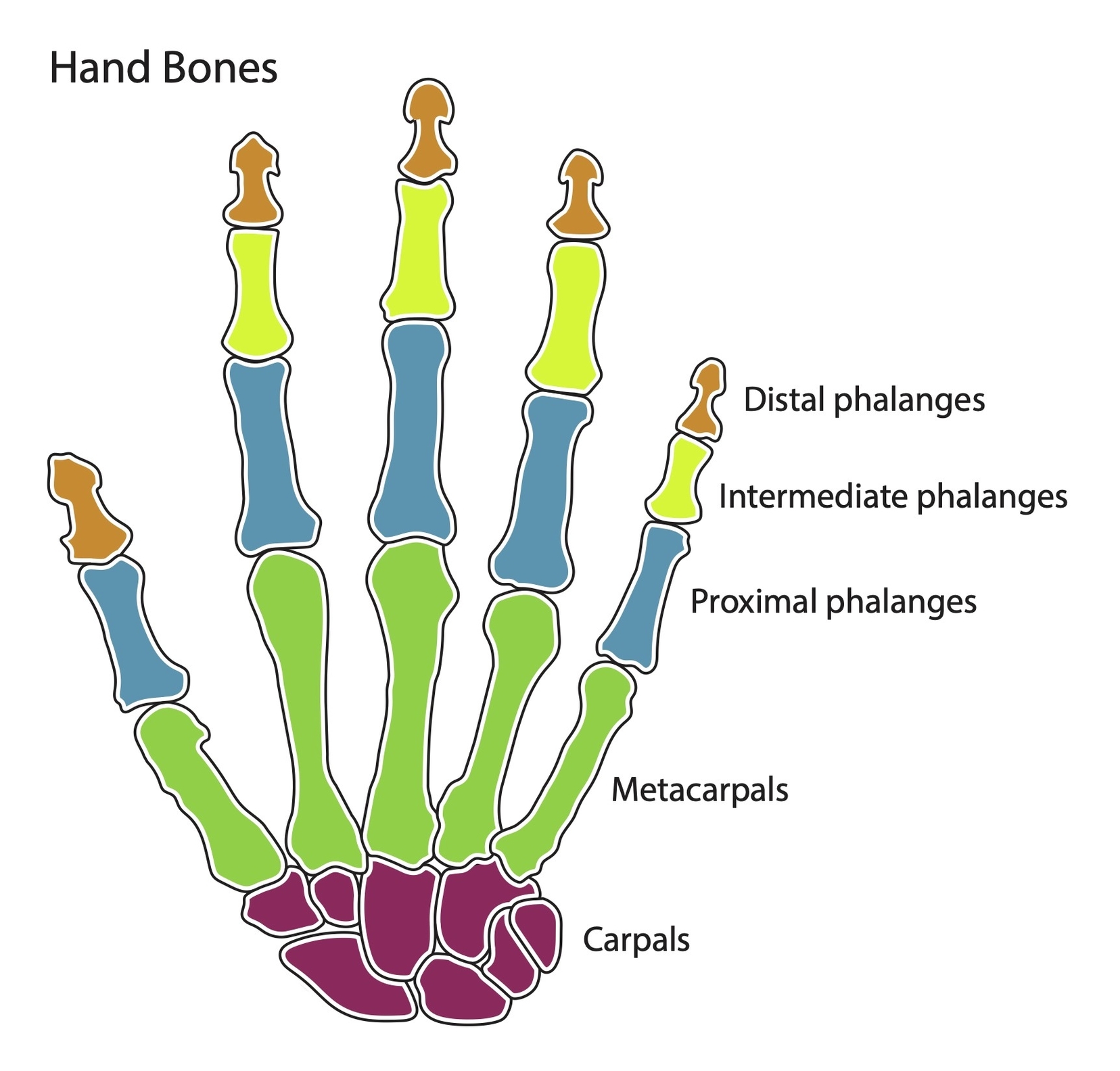
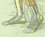
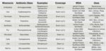
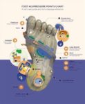
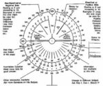
Skull Anatomy Coloring Pages

Skull Anatomy Coloring Pages
kull Anatomy Coloring Pages are educational tools that allow individuals to learn about the structure of the human skull through an interactive and creative activity. These coloring pages often depict the skull from various angles and highlight different components, providing a detailed view of the skull’s anatomy.

Types of Skull Coloring Pages

There are numerous types of skull coloring pages available, each offering a unique perspective or design:

1. Realistic Anatomy: These pages provide a detailed representation of the skull’s bone structure. They can be used for educational purposes, helping students understand the complex anatomy of the skull.

2. Abstract Designs: These pages feature skulls incorporated into abstract patterns or designs. They offer a more artistic approach to learning about the skull’s structure.

3. Cultural Illustrations: Some pages depict culturally significant skull designs, such as the sugar skulls associated with Dia de los Muertos.

4. Themed Skulls: These pages include skulls with various themes, such as pirate skulls, skulls with flower crowns, or skulls with butterfly wings.

Benefits of Skull Anatomy Coloring Pages
kull Anatomy Coloring Pages offer several benefits:

1. Educational Tool: They serve as an effective educational tool, especially for visual learners. By coloring the different parts of the skull, individuals can better understand its structure and function.

2. Artistic Expression: These pages provide an opportunity for artistic expression. Individuals can use various colors and techniques to personalize their skull anatomy coloring pages.

3. Relaxation and Stress Relief: Coloring is often seen as a relaxing activity. Focusing on the task can help reduce stress and promote mindfulness.

4. Accessibility: Many of these coloring pages are freely available online and can be downloaded and printed for use.

Conclusion
kull Anatomy Coloring Pages are a unique blend of art and science. They provide an engaging way to learn about the human skull’s structure while offering a platform for creativity and relaxation. Whether you’re interested in anatomy, looking for a relaxing activity, or seeking a fun way to learn, these coloring pages can be a great resource.

Skull Anatomy Coloring Pages Diagram - Skull Anatomy Coloring Pages Chart - Human anatomy diagrams and charts explained. This anatomy system diagram depicts Skull Anatomy Coloring Pages with parts and labels. Best diagram to help learn about health, human body and medicine.

Skull Anatomy Coloring Pages

Lower Leg Muscles And Tendons Anatomy

Lower Leg Muscles and Tendons Anatomy

The lower leg, located between the knee and ankle, is a complex structure comprising bones, muscles, tendons, nerves, and blood vessels. It plays a crucial role in weight-bearing activities such as walking, standing, running, and jumping.

Bones

The lower leg is supported by two strong, long bones: the tibia and the fibula. The tibia, or shinbone, is the main weight-bearing bone, located toward the middle of the lower leg. The fibula, or calf bone, is smaller and located on the outside of the lower leg.

Muscles

The lower leg is divided into four compartments that contain various muscles:

1. Anterior Compartment: This compartment, in front of the shin, holds the tibialis anterior, the extensor digitorum longus, the extensor hallucis longus, and the peroneus tertius muscles. These muscles pull the toes and feet upward, a process known as dorsiflexion.

2. Lateral Compartment: Located along the outside of the lower leg, it contains the peroneus longus and peroneus brevis muscles. These muscles pull the toes and feet outward and help with pointing the foot, or plantar flexion.

3. Posterior Compartment: This compartment holds the large muscles known as the calf muscles—the gastrocnemius and soleus. The gastrocnemius is shorter, thicker, and the most visible of the calf muscles. The soleus lies underneath. These muscles attach to the Achilles tendon and aid with plantar flexion.

4. Deep Posterior Compartment: The details of the muscles in this compartment are not provided in the search results.

Tendons

Tendons connect muscles to bones. When the muscle contracts, the tendons are pulled, and the bone is moved. The major tendon in the lower leg is the calcaneal tendon, or Achilles tendon. It attaches the muscles of the calf to the calcaneus. The action of the Achilles tendon allows for basic motions in the leg, such as walking and running.

Nerves

The lower leg is also home to nerve fibers, including the superficial fibular (or superficial peroneal) nerve, the deep fibular (or deep peroneal) nerve, and the tibial nerve.

Conclusion

The lower leg’s intricate anatomy enables it to perform key functions and withstand the body’s weight. Understanding its structure and function can help in diagnosing and treating common conditions that affect the lower leg, such as stress fractures, compartment syndrome, shin splints, and muscle tears.

Lower Leg Muscles And Tendons Anatomy Diagram - Lower Leg Muscles And Tendons Anatomy Chart - Human anatomy diagrams and charts explained. This anatomy system diagram depicts Lower Leg Muscles And Tendons Anatomy with parts and labels. Best diagram to help learn about health, human body and medicine.

Lower Leg Muscles And Tendons Anatomy

Brain Anatomy And Physiology Definition

Brain Anatomy and Physiology

The brain, a complex organ, is the central hub of the nervous system, controlling thought, memory, emotion, touch, motor skills, vision, breathing, temperature, hunger, and every process that regulates our body. It integrates sensory information and directs motor responses.

Composition

Weighing about 3 pounds in the average adult, the brain is about 60% fat. The remaining 40% is a combination of water, protein, carbohydrates, and salts. It contains blood vessels and nerves, including neurons and glial cells.

Gray Matter and White Matter

Gray and white matter are two different regions of the central nervous system. In the brain, gray matter refers to the darker, outer portion, while white matter describes the lighter, inner section underneath. Gray matter is primarily composed of neuron somas (the round central cell bodies), and white matter is mostly made of axons (the long stems that connect neurons together) wrapped in myelin (a protective coating). Gray matter is primarily responsible for processing and interpreting information, while white matter transmits that information to other parts of the nervous system.

Functioning

The brain sends and receives chemical and electrical signals throughout the body. Different signals control different processes, and your brain interprets each. Some make you feel tired, for example, while others make you feel pain. Some messages are kept within the brain, while others are relayed through the spine and across the body’s vast network of nerves to distant extremities.

Main Parts of the Brain

At a high level, the brain can be divided into the cerebrum, brainstem, and cerebellum.

– Cerebrum: The cerebrum (front of brain) comprises gray matter (the cerebral cortex) and white matter at its center. The largest part of the brain, the cerebrum initiates and coordinates movement and regulates temperature. Other areas of the cerebrum enable speech, judgment, thinking and reasoning, problem-solving, emotions, and learning.

– Cerebral Cortex: The cortex has a large surface area due to its folds, and comprises about half of the brain’s weight. The cerebral cortex is divided into two halves, or hemispheres. It is covered with ridges (gyri) and folds (sulci).

– Brainstem and Cerebellum: The brainstem and cerebellum control both voluntary movements, such as those involved in walking and in speech, and involuntary movements, such as breathing and reflex actions.

In conclusion, the brain is a marvel of nature, a complex organ that not only controls our bodily functions but also makes us who we are, shaping our thoughts, emotions, and behaviors..

Brain Anatomy And Physiology Definition Diagram - Brain Anatomy And Physiology Definition Chart - Human anatomy diagrams and charts explained. This anatomy system diagram depicts Brain Anatomy And Physiology Definition with parts and labels. Best diagram to help learn about health, human body and medicine.

Brain Anatomy And Physiology Definition

Human Abdomen Organs With Highlighted Stomach Examined

The human abdomen is a complex structure housing several vital organs. Let’s examine these organs, with a special focus on the stomach.

1. Stomach: Located in the upper part of the abdomen, the stomach plays a crucial role in digestion. It receives food from the esophagus and breaks it down both mechanically and chemically. The stomach’s three layers – the oblique layer, the middle circular layer, and the external longitudinal layer – work together to churn food. This mechanical breakdown is complemented by chemical digestion through stomach acids, including hydrochloric acid. The stomach also stores food until it’s ready to move further along the digestive tract.

2. Liver: Situated at the top of the abdominal cavity, the liver is the body’s largest organ. It acts as a filtration system, eliminating toxins and producing bile, which aids in the digestion and absorption of fats and fat-soluble vitamins.

3. Gallbladder: This small sac beneath the liver stores extra bile produced by the liver until it’s needed in the small intestine. Bile is crucial for digesting fats, excreting cholesterol, and even has antimicrobial activity.

4. Pancreas: This gland produces enzymes that help your body digest proteins, carbohydrates, and fats. It also makes hormones that help regulate the distribution of nutrients, including sugar.

5. Small Intestine: Occupying most of the abdominal cavity, this 21-foot long tube is where the majority of digestion occurs. It breaks down fats, starches, and proteins into fatty acids, which can then be absorbed.

6. Large Intestine: Despite its name, the large intestine is shorter than the small intestine but larger in girth. It’s the last part of the digestive tract and is made up of the cecum, colon, and rectum.

7. Kidneys: Positioned behind the intestines, the kidneys contain an estimated 1 million filtering units called nephrons. They play a vital role in processing the blood before it goes into general circulation.

8. Adrenal Glands: Located on top of the kidneys, these glands synthesize and secrete different sets of hormones. These hormones help the kidneys conserve sodium and water, and also support the body’s sexual functions.

9. Ureters: These two tubes carry urine from the kidneys to the urinary bladder.

10. Ribs: The main bones in the abdominal region, the ribs protect vital internal organs.

In conclusion, the human abdomen is a marvel of biological engineering, with each organ playing a unique and vital role in maintaining the body’s overall health and functionality. The stomach, in particular, serves as a critical junction in the digestive process, preparing food for further digestion and absorption in the intestines..

Human Abdomen Organs With Highlighted Stomach Examined Diagram - Human Abdomen Organs With Highlighted Stomach Examined Chart - Human anatomy diagrams and charts explained. This anatomy system diagram depicts Human Abdomen Organs With Highlighted Stomach Examined with parts and labels. Best diagram to help learn about health, human body and medicine.

Human Abdomen Organs With Highlighted Stomach Examined

Ear Anatomy And Sinus Connection

Ear Anatomy and Sinus Connection

The human ear is a complex sensory organ responsible for hearing and balance. It is anatomically divided into three parts: the external ear, the middle ear, and the internal ear.

External Ear
The external ear, like the middle ear, serves only to conduct sound to the inner ear. It consists of the auricle and external acoustic meatus (or ear canal).

Middle Ear
The middle ear contains the tympanic cavity, auditory ossicles, and muscles of the ossicles. Its function is to transform a high-amplitude low-force sound wave into a low-amplitude high-force vibration and transmit it to the internal ear.

Internal Ear
The internal ear comprises the bony labyrinth (vestibule, semicircular canals, cochlea) and membranous labyrinth (utricle, saccule, semicircular ducts, cochlear duct). The bony labyrinth supports its membranous counterparts, while the utricle and saccule provide information about the position of the head. The semicircular ducts provide information about movements of the head, and the cochlear duct provides hearing information.
inus Connection
The ear, nose, and throat are part of the upper respiratory system and share the same mucous membranes. The interconnected nature of these organs means that a disturbance in one can cause a problem in the others. The Eustachian tubes, which connect the middle ear to the nasopharynx, play a crucial role in this connection. These tubes are lined with mucous, just like the inside of the nose and throat.

The Eustachian tubes have several important functions, including allowing the passage of air from the ear to the sinuses, balancing pressure in the ear, draining excess fluid from the ear, and protecting the ears from hearing sounds from within the body.

The paranasal sinuses drain into the nasal passages at different points. The fluid trapped within the sinuses can travel to the ear through the Eustachian tubes. This interconnected system helps us breathe, smell, taste, and plays a defining role in our looks.

In conclusion, the ear’s anatomy and its connection to the sinuses is a complex and intricate system that allows us to hear and maintain balance, while also facilitating other crucial functions such as breathing and tasting. Any disturbance in this system can lead to various ailments, highlighting the importance of maintaining the health of our ears and sinuses.

Ear Anatomy And Sinus Connection Diagram - Ear Anatomy And Sinus Connection Chart - Human anatomy diagrams and charts explained. This anatomy system diagram depicts Ear Anatomy And Sinus Connection with parts and labels. Best diagram to help learn about health, human body and medicine.

Ear Anatomy And Sinus Connection

Internal Structure Of Human Stomach Visual

The human stomach, a key organ in the digestive system, is a complex structure with several distinct regions and layers. It plays a crucial role in the digestion of food and absorption of nutrients.

Location and Structure

The stomach is located in the upper abdomen on the left side of the body. It is a J-shaped organ that spans the region between the cardiac and pyloric orifices of the gastrointestinal tract. The stomach’s convex lateral surface is known as the greater curvature, while the concave medial border is the lesser curvature.

Parts of the Stomach

The stomach comprises four major regions:
1. Cardia: The area around the opening where the esophagus connects to the stomach.
2. Fundus: The dome-shaped part located to the left of the cardia.
3. Body: The main, central region of the stomach.
4. Pylorus: The lower part of the stomach that connects to the duodenum.

Layers of the Stomach

The stomach wall consists of several layers:
1. Mucosa: The innermost layer, which produces enzymes and acids for digestion.
2. Submucosa: Contains connective tissue, blood vessels, lymph vessels, and nerve cells.
3. Muscularis Externa: The primary muscle of the stomach, responsible for churning and mixing food.
4. Serosa: The outermost layer, a membrane that covers the stomach.

Function of the Stomach

The stomach’s primary function is to digest food and send it to the small intestine. It temporarily stores food, contracts and relaxes to mix and break down food, and produces enzymes and other specialized cells to digest food. The stomach works in conjunction with the rest of the gastrointestinal tract to break down food and liquid, absorb nutrients and water, and expel waste products of digestion through the large intestine.

Blood Supply and Innervation

The stomach receives its blood supply mainly from the celiac trunk. Innervation is provided via the vagus nerves and the celiac plexus.

Microscopic Anatomy

The inner part of the stomach lining, the gastric mucosa, consists of an outer layer of column-shaped cells, a lamina propria, and a thin layer of smooth muscle called the muscularis mucosa. Beneath the mucosa lies the submucosa, consisting of fibrous connective tissue.

In conclusion, the human stomach is a complex organ with a detailed internal structure. Its various parts and layers work together to perform the essential function of digesting food and absorbing nutrients, making it a vital component of the human digestive system..

Internal Structure Of Human Stomach Visual Diagram - Internal Structure Of Human Stomach Visual Chart - Human anatomy diagrams and charts explained. This anatomy system diagram depicts Internal Structure Of Human Stomach Visual with parts and labels. Best diagram to help learn about health, human body and medicine.

Internal Structure Of Human Stomach Visual

Virus Cell Structure

Virus Cell Structure

A virus is a tiny, infectious particle that can reproduce only by infecting a host cell. Viruses are much smaller than bacteria and consist of a single- or double-stranded nucleic acid (DNA or RNA) surrounded by a protein shell called a capsid. Some viruses also have an outer envelope composed of lipids and proteins.

Key Components of a Virus

1. Nucleic Acid Genome: A virus is made up of a DNA or RNA genome. This genome is the genetic material of the virus and contains the information needed for the virus to replicate.

2. Protein Capsid: The genome is enclosed within a protective protein coat called a capsid. The capsid is made from proteins that are encoded by viral genes within their genome. The shape of the capsid may vary from one type of virus to another.

3. Lipid Envelope: Many animal viruses also contain a lipid envelope. The envelope is made from portions of the host’s cell membrane. It surrounds the capsid and helps protect the virus from the host’s immune system. The envelope may also have receptor molecules that can bind with host cells, making it easier for the virus to infect the cells.

Types of Viruses Based on Structure

1. Helical Viruses: Helical capsids are made up of a single type of protein subunit stacked around a central axis to form a helical structure. This arrangement results in rod-shaped or filamentous virions. An example of a helical virus is the tobacco mosaic virus.

2. Icosahedral Viruses: Icosahedral capsid symmetry gives viruses a spherical appearance at low magnification, but the protein subunits are actually arranged in a regular geometrical pattern. They are not truly spherical.

Virus Reproduction

Viruses reproduce by infecting their host cells and reprogramming them to become virus-making “factories”. They “commandeer” the host cell and use its resources to make more viruses.

Conclusion

Viruses are unique entities that straddle the line between living and non-l

Virus Cell Structure Diagram - Virus Cell Structure Chart - Human anatomy diagrams and charts explained. This anatomy system diagram depicts Virus Cell Structure with parts and labels. Best diagram to help learn about health, human body and medicine.

Virus Cell Structure

Acupressure Points On Feet Charts

acupressure points on the feet. These points, deeply rooted in ancient healing practices, offer a pathway to well-being and balance. Without further ado, let’s explore their significance and benefits.

## What Is Reflexology?
Reflexology is a holistic practice that involves stimulating specific points on the feet. These points correspond to various organs and systems within the body. According to traditional Chinese medicine (TCM), energy flows through meridians, and these foot points serve as gateways to influence that energy. Here’s what you need to know:

1. Meridian Points: Imagine a map on the soles of your feet, each point representing an organ or body part. By applying pressure to these points, you can promote relaxation, alleviate pain, and enhance overall well-being.

2. Ancient Origins: Reflexology has ancient roots, dating back to 2330 B.C. Its name emerged in the United States during the early 1900s, but its practice spans cultures and centuries.

## Benefits of Foot Reflexology:
Let’s explore the remarkable benefits associated with acupressure points on the feet:

### 1. Stress and Relaxation:
– Reflexology is a powerful stress management tool. By applying pressure to specific foot points, blood flow increases throughout the body.
– Reduced stress and anxiety lead to relaxation. When you feel less tense, your entire system can unwind.

### 2. Pain Management:
– Reflexology isn’t just about relaxation; it also helps manage pain.
– Whether you’re dealing with acute discomfort or chronic conditions, foot reflexology may provide relief.
– By targeting specific points, you can ease pain and promote healing.

### 3. Digestion Enhancement:
– Certain foot points correspond to the digestive system.
– Stimulating these points may aid digestion, alleviate bloating, and improve overall gut health.

### 4. Eyestrain Relief:
– The eyes have their own reflexology points on the feet.
– If you spend hours staring at screens, consider giving these points some attention.

### 5. Improved Sleep:
– Insomnia and disrupted sleep patterns affect many.
– Reflexology may help regulate sleep cycles by promoting relaxation and balancing energy flow.

## Key Pressure Points on the Feet:
Now, let’s explore some of the main pressure points:

1. Ball of the Foot: This area corresponds to the chest. Applying pressure here may benefit respiratory health.

2. Arch Area: Linked to the abdomen, this point can aid digestion and alleviate stomach discomfort.

3. Heel: The heel connects to the pelvic area. It’s essential for overall balance and stability.

4. Outer Side of the Foot: This side influences arms, elbows, knees, and legs.

5. Inner Side of the Foot: Here, you’ll find points related to the neck, brain stem, thymus, spine, and bladder.

6. Outer Side of the Ankle: This area affects the lower back.

7. Inner Side of the Ankle: Lymph glands, fallopian tubes, and groin points reside here.

8. Top and Middle Area of the Foot: These points connect to the lungs, chest, breasts, and upper back.

Remember, reflexology is a journey. Regular sessions yield optimal benefits. So, kick off your shoes, explore these foot pathways, and embrace the ancient wisdom that lies beneath your soles. ????

For more detailed information, you can explore the [Healthline article](^1^) on foot reflexology..
o

Acupressure Points On Feet Charts Diagram - Acupressure Points On Feet Charts Chart - Human anatomy diagrams and charts explained. This anatomy system diagram depicts Acupressure Points On Feet Charts with parts and labels. Best diagram to help learn about health, human body and medicine.

Acupressure Points On Feet Charts

Human Anatomy For Muscle, Reproductive, And Skeleton Represented

Human Anatomy: Muscle, Reproductive, and Skeletal Systems

Muscular System

The muscular system is responsible for movement, posture, and balance. It consists of three types of muscle tissue: skeletal, smooth, and cardiac. Skeletal muscles, numbering over 600, create movement in the body. They work in groups to move the skeleton, with movements being nearly involuntary, yet requiring conscious effort. Cardiac muscle, found in the heart, is responsible for the rhythmic contractions that pump blood throughout the body. Smooth muscle, found in the walls of hollow organs and blood vessels, propels substances through the body.

Reproductive System

The human reproductive system allows for the production and fertilization of gametes, leading to the creation of offspring[^10^]. In males, the testes produce sperm, and the penis delivers the sperm for potential fertilization[^10^]. In females, the ovaries produce eggs, the uterus houses the developing fetus, and the breasts produce milk for the newborn. The reproductive process involves the release of an egg, internal fertilization by sperm, transport of the fertilized egg to the uterus, implantation in the uterine wall, gestation, birth, and postnatal care[^10^].
keletal System

The skeletal system serves as the body’s framework, providing support, protection, and enabling motion?. It consists of 206 bones, divided into the axial and appendicular skeletons?. The axial skeleton includes the vertebral column and much of the skull, providing support and protection for the body’s central parts?. The appendicular skeleton includes the pelvic and pectoral girdles and the bones of the limbs?. The skeletal system also includes ligaments, which attach bone to bone, and cartilage, which provides padding between bones?.

In conclusion, the muscular, reproductive, and skeletal systems each play crucial roles in the human body. The muscular system enables movement and maintains posture, the reproductive system allows for the continuation of human species, and the skeletal system provides structural support and protection for the body’s organs. Each system, with its unique structure and function, contributes to the overall health and well-being of an individual..

Human Anatomy For Muscle, Reproductive, And Skeleton Represented Diagram - Human Anatomy For Muscle, Reproductive, And Skeleton Represented Chart - Human anatomy diagrams and charts explained. This anatomy system diagram depicts Human Anatomy For Muscle, Reproductive, And Skeleton Represented with parts and labels. Best diagram to help learn about health, human body and medicine.

Human Anatomy For Muscle, Reproductive, And Skeleton Represented

Hamstring Muscleshamstring Origin Anatomy

The Hamstring Muscles are a group of three muscles located in the posterior compartment of the thigh. These muscles include the Semitendinosus, Semimembranosus, and Biceps Femoris.
emitendinosus
– Origin: The lower medial facet of the lateral section of the ischial tuberosity.
– Insertion: A vertical line on the medial surface of the medial condyle of the tibia just behind the insertion of sartorius and behind and below the attachment of gracilis.
– Nerve: Tibial division of the sciatic nerve (L5, S1 and 2).
– Function: Hip extension, Knee flexion, Internal rotation of lower leg when the knee is flexed.
emimembranosus
– Origin: The upper lateral facet on the ischial tuberosity.
– Insertion: A horizontal groove on the posteromedial surface of the medial tibial condyle.
– Nerve: Tibial division of the sciatic nerve (L5, S1 and 2).
– Function: Hip extension, Knee flexion, Internal rotation of lower leg when the knee is flexed.

Biceps Femoris – Long Head
– Origin: The lower medial facet on the ischial tuberosity with the tendon of semitendinosus, spreading onto the sacrotuberous ligament.
– Insertion: The head of the fibular, the lateral tibial condyle and the posterior aspect of the lateral intermuscular septum.
– Nerve: Tibial division of the sciatic nerve (L5, S1 and 2).
– Function: Knee flexion, Hip extension, External rotation of lower leg when knee slightly flexed, Assist in external rotation of the thigh when hip extended.

Biceps Femoris – Short Head
– Origin: The lower half of the lateral lip of the linea aspera.
– Insertion: The head of the fibular, the lateral tibial condyle and the posterior aspect of the lateral intermuscular septum.
– Nerve: The common peroneal division of the sciatic nerve (L5, S1 and 2).
– Function: Knee flexion, External rotation of lower leg when knee slightly flexed.

The primary function of the hamstrings is to flex the knee joint and extend the hip, enabling some of the essential lower limb activities such as walking, running, and climbing. The hamstrings have an important stabilizing function as well; they are inactive when the bodyweight is equally distributed between both lower limbs in a standing position. However, when a person starts tilting forward, these muscles activate and counteract the tilting movement in order to stabilize the hip joint and prevent falling. Also, due to the location of their insertions, the hamstrings act together with the collateral ligaments to stabilize the knee joint..

Hamstring Muscleshamstring Origin Anatomy Diagram - Hamstring Muscleshamstring Origin Anatomy Chart - Human anatomy diagrams and charts explained. This anatomy system diagram depicts Hamstring Muscleshamstring Origin Anatomy with parts and labels. Best diagram to help learn about health, human body and medicine.

Hamstring Muscleshamstring Origin Anatomy

Bones Of The Foot And Ankle

The foot and ankle form a complex system which consists of 28 bones, 33 joints, 112 ligaments, controlled by 13 extrinsic and 21 intrinsic muscles?. The foot is subdivided into the rearfoot, midfoot, and forefoot?.

Ankle Anatomy

The ankle joint, also known as the talocrural joint, allows dorsiflexion and plantar flexion of the foot. It is made up of three joints: upper ankle joint (tibiotarsal), talocalcaneonavicular, and subtalar joints. The last two together are called the lower ankle joint. The upper ankle joint is formed by the inferior surfaces of tibia and fibula, and the superior surface of talus. The lower ankle joint is formed by the talus, calcaneus, and navicular bone.

Bones of the Foot

The foot begins at the lower end of the tibia and fibula, the two bones of the lower leg. At the base of those, a grouping of bones form the tarsals, which make up the ankle and upper portion of the foot. The bones of the foot are organized into the tarsal bones, metatarsal bones, and phalanges.

1. Tarsal Bones: These include the talus and calcaneus, which form the ankle joint and the heel, and the navicular, cuboid, and three cuneiforms, which form the arch of the foot.
2. Metatarsal Bones: These are the long bones that connect the tarsals to the toes.
3. Phalanges: These are the small bones that form the toes. The great toe consists of two phalanges (proximal, distal), while the remaining four toes have three phalanges (proximal, middle, distal).

Joints of the Foot

The foot has several joints, including intertarsal, tarsometatarsal, metatarsophalangeal, and interphalangeal joints. The great toe has one interphalangeal joint, while the other four toes have two (proximal, distal) interphalangeal joints.

Muscles of the Foot

The foot has several muscles that allow movements like foot inversion, foot eversion, toe flexion, toe extension, toe abduction, and toe adduction. These include dorsal muscles (extensor digitorum brevis, extensor hallucis brevis), lateral plantar muscles (abductor digiti minimi, flexor digiti minimi brevis, opponens digiti minimi), central plantar muscles (flexor digitorum brevis, quadratus plantae, lumbricals, plantar interossei, dorsal interossei), and medial plantar muscles (abductor hallucis, adductor hallucis, flexor hallucis brevis).

In conclusion, the foot and ankle’s complex structure, consisting of numerous bones, joints, and muscles, allows for a wide range of movements and provides the necessary stability and flexibility for various activities such as walking, running, and jumping..

Bones Of The Foot And Ankle Diagram - Bones Of The Foot And Ankle Chart - Human anatomy diagrams and charts explained. This anatomy system diagram depicts Bones Of The Foot And Ankle with parts and labels. Best diagram to help learn about health, human body and medicine.

Bones Of The Foot And Ankle

Human Leg Muscles With Labels

The human leg, a marvel of biological engineering, is powered by a complex network of muscles. These muscles, working in harmony, enable us to perform a wide range of movements, from walking and running to jumping and standing on our toes.

Upper Leg Muscles

The upper leg, or thigh, houses some of the body’s strongest muscles, including the quadriceps and hamstrings.

1. Quadriceps: These are the major extensors of the knee and are the strongest and leanest muscles in the body. They consist of four muscles:
– Vastus lateralis: The largest of the quadriceps, it extends from the top of the femur to the kneecap.
– Vastus medialis: A teardrop-shaped muscle of the inner thigh that attaches along the femur and down to the inner border of the kneecap.
– Vastus intermedius: Located between the vastus medialis and the vastus lateralis, it is the deepest of the quadriceps muscles.
– Rectus femoris: This muscle attaches to the kneecap and has the least effect on flexion of the knee among the quadriceps muscles.

2. Hamstrings: These are three muscles at the back of the thigh that affect hip and knee movement.
– Biceps femoris: This long muscle flexes the knee.
– Semimembranosus: This long muscle extends from the pelvis to the tibia, extending the thigh, flexing the knee, and helping rotate the tibia.
– Semitendinosus: This muscle also extends the thigh and flexes the knee.

Lower Leg Muscles

The lower leg muscles, including the calf muscles, work with other muscles to help move the feet.

1. Gastrocnemius (calf muscle): One of the large muscles of the leg, it connects to the heel and flexes and extends the foot, ankle, and knee.
2. Soleus: This muscle extends from the back of the knee to the heel and is important in walking and standing.
3. Plantaris: This small, thin muscle is absent in about 10 percent of people. The gastrocnemius muscle supersedes its function.

Achilles Tendon

Possibly the most important tendon in terms of mobility is the Achilles tendon. This important tendon in the back of the calf and ankle connects the plantaris, gastrocnemius, and soleus muscles to the heel bone. It stores the elastic energy needed for running, jumping, and other physical activity.

In conclusion, the muscles of the human leg, from the powerful quadriceps and hamstrings to the pivotal calf muscles, play a crucial role in our mobility. They not only enable movement but also provide stability and support, making them integral to our daily lives.

Human Leg Muscles With Labels Diagram - Human Leg Muscles With Labels Chart - Human anatomy diagrams and charts explained. This anatomy system diagram depicts Human Leg Muscles With Labels with parts and labels. Best diagram to help learn about health, human body and medicine.

Human Leg Muscles With Labels

Human Body Tissues Explained

Human Body Tissues Explained

The human body is a complex structure composed of four primary tissue types: epithelial, connective, muscle, and nervous. These tissues, each with their unique structure and function, work together to maintain the overall health and functionality of the body.

1. Epithelial Tissue: This tissue type covers the exterior surfaces of the body, lines internal cavities and passageways, and forms certain glands. It is made of layers of cells that cover the surfaces of the body that come into contact with the exterior world.

2. Connective Tissue: As its name implies, connective tissue binds the cells and organs of the body together. It provides support and structure to the body and is essential for our overall health and well-being.

3. Muscle Tissue: Muscle tissue contracts forcefully when excited, providing movement. It is crucial for our ability to move and perform physical tasks.

4. Nervous Tissue: Nervous tissue is excitable, allowing for the generation and propagation of electrochemical signals in the form of nerve impulses that communicate between different regions of the body. It is essential for our ability to sense and respond to our environment.

These primary tissue types originate from the three germ layers formed during embryonic development: ectoderm (outer), mesoderm (middle), and endoderm (inner). The cells composing a tissue share a common embryonic origin.

The organization of these tissues into organs and organ systems allows the body to function as a whole. For example, the digestive system is responsible for taking in and processing food, while the respiratory system—working with the circulatory system—is responsible for taking up oxygen and getting rid of carbon dioxide. The muscular and skeletal systems are crucial for movement; the reproductive system handles reproduction; and the excretory system gets rid of metabolic waste.

In conclusion, understanding the various primary tissue types present in the human body is essential for understanding the structure and function of organs which are composed of two or more primary tissue types. The survival of the organism depends on the integrated activity of all the organ systems, often coordinated by the endocrine and nervous systems..

Human Body Tissues Explained Diagram - Human Body Tissues Explained Chart - Human anatomy diagrams and charts explained. This anatomy system diagram depicts Human Body Tissues Explained with parts and labels. Best diagram to help learn about health, human body and medicine.

Human Body Tissues Explained

Head And Neck Muscles Example Of

The muscles of the head and neck are intricate and diverse, responsible for a variety of crucial tasks such as movement of the head and neck, chewing and swallowing, speech, facial expressions, and movement of the eyes. These tasks require both strong, forceful movements and some of the fastest, finest, and most delicate adjustments in the entire human body.

Facial Muscles

The muscles of the face are unique among groups of muscles in the body. While most muscles connect to and move only bones, facial muscles mostly connect bones to skin. These muscles, including the zygomaticus major and orbicularis oris, pull on the skin to produce a seemingly infinite number of facial expressions and to move the lips and cheeks during speech and eating.

Muscles of Mastication

Producing the body’s ability to close the mouth, bite, and chew food, the muscles of mastication move the mandible relative to the rest of the skull. These muscles, including the masseter and temporalis, elevate the jaw forcefully during chewing and gently during speech.

Muscles of the Tongue

An extensive complement of tightly interlaced muscles allows the tongue a range of complex movements for chewing and swallowing, as well as the important function of producing speech. Of these, four extrinsic muscle sets (connecting the tongue to the surrounding bones) move the tongue in virtually any direction, with fine shape changes (such as for speech) the province of the four intrinsic tongue muscles.

Muscles of the Eye
ix extrinsic eye muscles provide superior, inferior, lateral, and medial motion, as well as rotation of the eyeball. These muscles produce extremely fine movements almost constantly throughout the day with tremendous speed and accuracy.

Neck Muscles

The muscles of the neck are mainly responsible for the movement of the head in all directions. They consist of 3 main groups of muscles: anterior, lateral, and posterior groups, based on their position in the neck. The position of a muscle or group of muscles in the neck generally relates to the function of the muscles. For example, the muscles in the posterior neck are responsible for extension of the neck.

Anterior Neck Muscles

The anterior neck muscles are a group of muscles covering the anterior aspect of the neck. They are further divided into 3 subgroups: The superficial muscles are the most superficial in the anterior neck, and include the platysma and sternocleidomastoid. The suprahyoid muscles, as the name suggests, are found superior to the hyoid bone, and include the digastric, mylohyoid, geniohyoid and stylohyoid.

Posterior Neck Muscles

Posterior neck muscles include the splenius capitis and splenius cervicis, strap-like muscles in the back of your neck that help you extend and rotate your head. Suboccipital muscles are four muscles just below the occipital bone at the base of your skull. They help extend your head in different directions.

In conclusion, the muscles of the head and neck are complex and diverse, each serving a specific function that contributes to our daily activities. Understanding these muscles and their functions can provide insight into how we perform such tasks as speaking, eating, expressing emotions, and moving our heads.

Head And Neck Muscles Example Of Diagram - Head And Neck Muscles Example Of Chart - Human anatomy diagrams and charts explained. This anatomy system diagram depicts Head And Neck Muscles Example Of with parts and labels. Best diagram to help learn about health, human body and medicine.

Head And Neck Muscles Example Of

How Did The Apostles Die

The Apostles, also known as the Twelve, were the primary disciples of Jesus Christ and played a crucial role in spreading Christianity throughout the ancient world. Many of them faced persecution and martyrdom as a direct result of their ministry efforts. The New Testament records the deaths of only two apostles, James and Judas Iscariot. For the others, we rely on historical information and traditions, which often present multiple accounts of their deaths.

1. Peter: Tradition claims that Peter died during Nero’s persecution of the Christians around A.D. 64-68. He was crucified upside down on a cross, allegedly because he didn’t consider himself worthy of dying the same death as Jesus.

2. Andrew: Andrew was crucified on a cross shaped like an “X”, now known as the St. Andrew’s cross. He was tied, not nailed, to the cross, and it took several days before he died. It is said that he preached while hanging on the cross.

3. James: James, the son of Zebedee, was beheaded by King Herod I around A.D. 44, launching a new persecution of Christians. He was the first martyr among the twelve apostles.

4. John: One tradition says John was on the Isle of Patmos before he was released and went to Ephesus, where he died around A.D. 100-105.

The Bible does not report how all of the apostles died, so we cannot be confident about some of them. It’s commonly believed that only one apostle, John, died of natural causes. However, some accounts suggest there may have

How Did The Apostles Die Diagram - How Did The Apostles Die Chart - Human anatomy diagrams and charts explained. This anatomy system diagram depicts How Did The Apostles Die with parts and labels. Best diagram to help learn about health, human body and medicine.

How Did The Apostles Die

Human Anatomy For Muscle, Reproductive, And Skeleton

Human Anatomy: Muscle, Reproductive, and Skeletal Systems

Muscular System
The muscular system is responsible for movement, posture, and balance. It consists of three types of muscles: skeletal, smooth, and cardiac.

– Skeletal Muscles: These muscles are attached to the bones by tendons and work in groups to move the skeleton. They make up about 40% of a person’s body weight.
– Smooth Muscles: Found in the walls of hollow organs, blood vessels, and respiratory passageways, they contract in response to stimuli and nerve impulses.
– Cardiac Muscles: These muscles make up the walls of the heart and are responsible for the rhythmic contractions that pump blood through the body.

Reproductive System
The reproductive system is responsible for human reproduction and bearing live offspring?. It includes both internal and external genitalia?.

– Male Reproductive System: The male reproductive system includes the testes that produce sperm and a penis for delivery?. The sperm mature in the testes and then enter the epididymis for further maturation?.
– Female Reproductive System: The female reproductive system includes the ovaries that produce eggs, a uterus for baby development, and breasts for milk production?. The ovum is released at a specific time in the reproductive cycle for internal fertilization by sperm cells?.
keletal System
The skeletal system serves as a framework for the body, providing shape, stability, and protection of internal organs. It consists of 206 bones, ligaments, and cartilages.

– Axial Skeleton: This includes the vertebral column (the spine) and much of the skull, providing the main support of the trunk.
– Appendicular Skeleton: This includes the pelvic (hip) and pectoral (shoulder) girdles and the bones and cartilages of the limbs.

In conclusion, the muscular, reproductive, and skeletal systems play crucial roles in the functioning of the human body. Each system has a unique structure and specific role, contributing to our movement, reproduction, and structural support.

Human Anatomy For Muscle, Reproductive, And Skeleton Diagram - Human Anatomy For Muscle, Reproductive, And Skeleton Chart - Human anatomy diagrams and charts explained. This anatomy system diagram depicts Human Anatomy For Muscle, Reproductive, And Skeleton with parts and labels. Best diagram to help learn about health, human body and medicine.

Human Anatomy For Muscle, Reproductive, And Skeleton

Human Leg Muscle Anatomy Examined

The human leg, a complex structure with numerous muscles, plays a pivotal role in body movement and support. The majority of leg muscles are considered long muscles, stretching great distances to move skeletal bones and facilitate body movement.

Upper Leg Muscles

The upper leg comprises the quadriceps and hamstrings. The quadriceps, the body’s strongest and leanest muscles, are major extensors of the knee. They include:

1. Vastus lateralis: The largest of the quadriceps, it extends from the top of the femur to the kneecap.
2. Vastus medialis: A teardrop-shaped muscle of the inner thigh, it attaches along the femur and down to the inner border of the kneecap.
3. Vastus intermedius: Located between the vastus medialis and the vastus lateralis at the front of the femur, it is the deepest of the quadriceps muscles.
4. Rectus femoris: This muscle attaches to the kneecap.

The hamstrings, three muscles at the back of the thigh, affect hip and knee movement. They include:

1. Biceps femoris: This long muscle flexes the knee.
2. Semimembranosus: This long muscle extends from the pelvis to the tibia.
3. Semitendinosus: This muscle extends the thigh and flexes the knee.

Lower Leg Muscles

The lower leg muscles, supported by the fibula and the tibia (shinbone), are pivotal to movement of the ankle, foot, and toes. Some of the major muscles of the calf include:

1. Gastrocnemius (calf muscle): One of the large muscles of the leg, it connects to the heel. It flexes and extends the foot, ankle, and knee.
2. Soleus: This muscle extends from the back of the knee to the heel. It is important in walking and standing.
3. Plantaris: This small, thin muscle is absent in about 10 percent of people. The gastrocnemius muscle supersedes its function.

The Achilles tendon, connecting the plantaris, gastrocnemius, and soleus muscles to the heel bone, stores the elastic energy needed for running, jumping, and other physical activity.

Functional Groups

The leg muscles are organized into three groups: anterior (dorsiflexor) group, posterior (plantar flexor) group, and lateral (fibular) group. These groups produce different movements in the ankle and foot, crucial for activities such as walking, running, and dancing.

The anterior group, including the tibialis anterior, extensor digitorum longus, fibularis tertius, and extensor hallucis longus, primarily produces dorsiflexion of the foot at the ankle joint. The posterior group, comprising the gastrocnemius, plantaris, soleus

Human Leg Muscle Anatomy Examined Diagram - Human Leg Muscle Anatomy Examined Chart - Human anatomy diagrams and charts explained. This anatomy system diagram depicts Human Leg Muscle Anatomy Examined with parts and labels. Best diagram to help learn about health, human body and medicine.

Human Leg Muscle Anatomy Examined

Human Earanatomy Of Human Ear

Anatomy of the Human Ear

The human ear is a complex organ that serves two primary functions: hearing and maintaining balance. It is anatomically divided into three parts: the outer ear, the middle ear, and the inner ear.

1. Outer Ear

The outer ear consists of the visible portion called the auricle or pinna, which projects from the side of the head, and the short external auditory canal. The inner end of this canal is closed by the tympanic membrane, commonly known as the eardrum. The function of the outer ear is to collect sound waves and guide them to the tympanic membrane.

2. Middle Ear

The middle ear is a narrow air-filled cavity in the temporal bone. It is spanned by a chain of three tiny bones — the malleus (hammer), incus (anvil), and stapes (stirrup), collectively known as the auditory ossicles. This ossicular chain conducts sound from the tympanic membrane to the inner ear.

3. Inner Ear

The inner ear, also known as the labyrinth, is a complicated system of fluid-filled passages and cavities located deep within the temporal bone. It consists of two functional units: the vestibular apparatus, which contains the sensory organs of postural equilibrium, and the cochlea, which contains the sensory organ of hearing. These sensory organs are highly specialized endings of the eighth cranial nerve, also known as the vestibulocochlear nerve.

Function of the Ear

The primary function of the ear is hearing, which involves the conversion of sound waves into electrochemical impulses, a process known as transduction. The ear also maintains the sense of balance or equilibrium. The vestibular apparatus provides information about the position and movements of the head, while the cochlea provides hearing information.

Clinical Relations

Various conditions can affect the ear, including otitis (inflammation of the ear), blockage of the auditory (Eustachian) tube, and high tone deafness. Understanding the anatomy of the ear is crucial for diagnosing and treating these conditions.

In conclusion, the human ear is a remarkable organ that plays a vital role in our ability to interact with our environment. Its complex structure enables it to perform its dual functions of hearing and maintaining balance, making it an essential part of our sensory system..

Human Earanatomy Of Human Ear Diagram - Human Earanatomy Of Human Ear Chart - Human anatomy diagrams and charts explained. This anatomy system diagram depicts Human Earanatomy Of Human Ear with parts and labels. Best diagram to help learn about health, human body and medicine.

Human Earanatomy Of Human Ear

External Anatomy Of The Left Human Eye

The human eye is a complex organ that allows us to perceive the world around us. The external anatomy of the eye includes several key components, each with a specific function.

1. Orbit: The eye sits in a protective bony socket called the orbit.

2. Extraocular Muscles: Six extraocular muscles in the orbit are attached to the eye. These muscles move the eye up and down, side to side, and rotate the eye.

3. Sclera: The extraocular muscles are attached to the white part of the eye called the sclera. This is a strong layer of tissue that covers nearly the entire surface of the eyeball.

4. Conjunctiva: The surface of the eye and the inner surface of the eyelids are covered with a clear membrane called the conjunctiva.

5. Tear Film: The layers of the tear film keep the front of the eye lubricated. Tears lubricate the eye and are made up of three layers. These three layers together are called the tear film.

6. Cornea: Light is focused into the eye through the clear, dome-shaped front portion of the eye called the cornea.

7. Anterior Chamber: Behind the cornea is a fluid-filled space called the anterior chamber. The fluid is called aqueous humor.

8. Iris and Pupil: Behind the anterior chamber is the eye’s iris (the colored part of the eye) and the dark hole in the middle called the pupil. Muscles in the iris dilate (widen) or constrict (narrow) the pupil to control the amount of light reaching the back of the eye.

9. Lens: Directly behind the pupil sits the lens. The lens focuses light toward the back of the eye. The lens changes shape to help the eye focus on objects up close.

10. Eyelids: The upper and lower eyelids form a moist region around the eye, and protect the surface of the eye from injury, infection, and disease.

11. Eyelashes: The eyelashes are the hairs that grow along the edges of the upper and lower eyelids. The eyelashes protect the eye from foreign particles, such as dust, pollen, and debris.

12. Meibomian Glands: Meibomian glands are the oil glands located inside the eyelids. Their opening pores line the edges of the eyelids, near the eyelashes.

Each of these components plays a crucial role in the overall function of the eye. They work together to protect the eye, control the amount of light that enters, and focus that light onto the retina, allowing us to see the world around us..

External Anatomy Of The Left Human Eye Diagram - External Anatomy Of The Left Human Eye Chart - Human anatomy diagrams and charts explained. This anatomy system diagram depicts External Anatomy Of The Left Human Eye with parts and labels. Best diagram to help learn about health, human body and medicine.

External Anatomy Of The Left Human Eye

Ear Anatomysimple Ear Anatomy Visual

Anatomy of the Human Ear

The human ear is a complex organ that serves two primary functions: hearing and maintaining balance. It is anatomically divided into three main parts: the outer ear, the middle ear, and the inner ear.

1. Outer Ear

The outer ear consists of the visible portion called the auricle, or pinna, and the external auditory canal. The auricle collects sound waves and guides them into the auditory canal. The inner end of the canal is closed by the tympanic membrane, commonly known as the eardrum. The function of the outer ear is to collect sound waves and guide them to the tympanic membrane.

2. Middle Ear

The middle ear is an air-filled cavity in the temporal bone. It contains a chain of three tiny bones — the malleus (hammer), incus (anvil), and stapes (stirrup), collectively known as the auditory ossicles. These bones conduct sound from the tympanic membrane to the inner ear.

3. Inner Ear

The inner ear, also known as the labyrinth, is a complex system of fluid-filled passages and cavities. It consists of two functional units: the vestibular apparatus and the cochlea. The vestibular apparatus, which includes the vestibule and semicircular canals, maintains balance. The cochlea, on the other hand, is responsible for hearing. These sensory organs are highly specialized endings of the eighth cranial nerve, also known as the vestibulocochlear nerve

Ear Anatomysimple Ear Anatomy Visual Diagram - Ear Anatomysimple Ear Anatomy Visual Chart - Human anatomy diagrams and charts explained. This anatomy system diagram depicts Ear Anatomysimple Ear Anatomy Visual with parts and labels. Best diagram to help learn about health, human body and medicine.

Ear Anatomysimple Ear Anatomy Visual