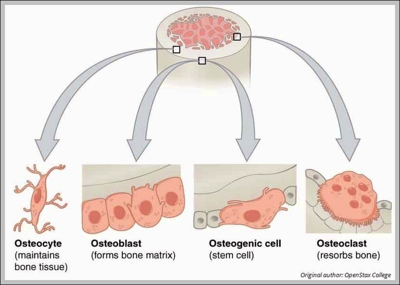
Cellular Components of Bone Diagram

Cellular Components of Bone Diagram

The Cellular Components of Bone diagram zooms to the microscopic level with osteoblasts building matrix, osteocytes living inside lacunae, osteoclasts breaking bone down, and the osteon units with their central…
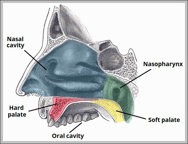
The Hard and Soft Palate Diagram

The Hard and Soft Palate Diagram

Hard palate (maxilla/palatine bones) forms anterior roof of mouth; soft palate (muscular: tensor/levator veli palatini, palatoglossus/pharyngeus) hangs posteriorly with uvula. It separates oral/nasal cavities during swallowing.
Aorta Diagram

Aorta Diagram

The aorta begins at left ventricle (ascending), arches leftward (arch with three branches), descends thoracic then abdominal to bifurcate at L4 into common iliacs. It supplies all systemic arteries except…
Types of Synovial Joint Diagram

Types of Synovial Joint Diagram

Types of synovial joints: plane (gliding, e.g., intercarpal), hinge (uniaxial, e.g., elbow), pivot (rotation, e.g., atlantoaxial), condyloid (biaxial, e.g., wrist), saddle (biaxial, e.g., thumb CMC), ball-and-socket (multiaxial, e.g., hip). All…
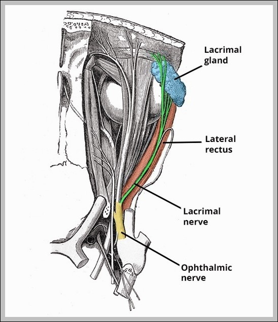
Course of the Lacrimal Nerve Diagram

Course of the Lacrimal Nerve Diagram

Lacrimal nerve (V1 branch) enters orbit via superior orbital fissure, runs superolateral, supplies lacrimal gland (parasympathetic hitchhike from facial via pterygopalatine ganglion), conjunctiva, upper eyelid skin. It communicates with zygomatic…
Anatomical Relations of the Prostate Gland Diagram

Anatomical Relations of the Prostate Gland Diagram

Anatomical relations of prostate gland: anterior pubic symphysis/vesicoprostatic space, posterior rectum (separated by DenonvilliersÂ’ fascia), superior bladder neck, inferior urogenital diaphragm, lateral levator ani. Seminal vesicles posterior-superior; urethra passes through.