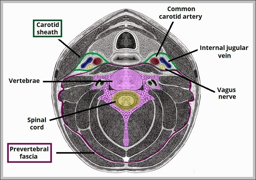
CPR Technique

CPR Technique

A CPR technique illustration demonstrates correct hand placement, compression depth, and rhythm. Sequential steps show how chest compressions and rescue breaths maintain circulation during cardiac arrest, emphasizing lifesaving timing and…
Digestion of Proteins Physiology

Digestion of Proteins Physiology

Digestion of proteins physiology involves not just enzymes but sophisticated regulation to match secretion to dietary intake, starting with the cephalic phase where sight or smell of food stimulates vagal…
Muscular and Elastic Artery Arteriole

Muscular and Elastic Artery Arteriole

Muscular arteries like femoral or radial distribute blood flow via thick tunica media of smooth muscle regulating resistance through vasoconstriction or dilation, elastic arteries like aorta buffer pulsatile flow with…
Vascular Homeostasis Flow Art

Vascular Homeostasis Flow Art

Vascular homeostasis maintains flow via autoregulation myogenic metabolic matching perfusion demand, endothelial nitric oxide prostacyclin vasodilation shear stress, endothelin angiotensin constriction, arteriolar tone dominant resistance site, capillary hydrostatic oncotic pressures…
FlowChart Veins into VenaCava

FlowChart Veins into VenaCava

Venous blood from the body converges into two great veins: the superior and inferior vena cava. Head and neck veins drain into the brachiocephalic veins, which merge into the superior…
Iliac Artery Branches Chart

Iliac Artery Branches Chart

The common iliac arteries branch from abdominal aorta bifurcation at L4, each dividing into external iliac supplying lower limb via femoral and internal iliac feeding pelvis. Internal branches include superior…
Fetal Circulatory System

Fetal Circulatory System

The fetal circulatory system is beautifully adapted to bypass the lungs and liver while maximizing placental exchange. Blood leaves the fetus through two umbilical arteries branching from the iliac arteries,…
Major Systemic Artery

Major Systemic Artery

Major systemic arteries begin with ascending aorta giving coronaries, arch branching brachiocephalic, left common carotid, left subclavian, then descending thoracic supplying intercostals, abdominal continuing as celiac, mesenteric, renal, gonadal, iliacs,…
Lower Limb Arteries Chart

Lower Limb Arteries Chart

The arterial supply to lower limbs forms a continuous chain with branches providing redundancy, starting with common femoral dividing into superficial femoral through adductor canal and profunda femoris laterally, then…
Comparison of Artery and Vein

Comparison of Artery and Vein

A side-by-side vascular comparison highlights the structural differences between arteries and veins. Thick muscular walls, narrow lumens, and elastic layers are shown in arteries, while veins appear thinner with wider…
Stethoscope Placement

Stethoscope Placement

Stethoscope placement for heart sounds uses four standard sites: aortic second right intercostal sternal border high-pitched semilunar closures, pulmonic second left for same, tricuspid lower left sternal for right AV,…
Cooperation Between Innate and Immune Responses

Cooperation Between Innate and Immune Responses

An immune system interaction diagram illustrates how innate and adaptive responses work together. Early defenses such as macrophages and inflammation are shown activating and guiding lymphocytes. This cooperation explains faster…
Embroyonic Disc Amniotic Cavity Yolk Sac

Embroyonic Disc Amniotic Cavity Yolk Sac

The embryonic disc with amniotic cavity and yolk sac represents the early post-implantation stage, where the bilaminar disc of epiblast and hypoblast forms, then gastrulation creates a trilaminar disc with…
Cardiac Cycle vs Heart Sounds

Cardiac Cycle vs Heart Sounds

A comparison of the cardiac cycle and heart sounds visually links mechanical events in the heart to what is heard through a stethoscope. The diagram aligns phases such as atrial…