
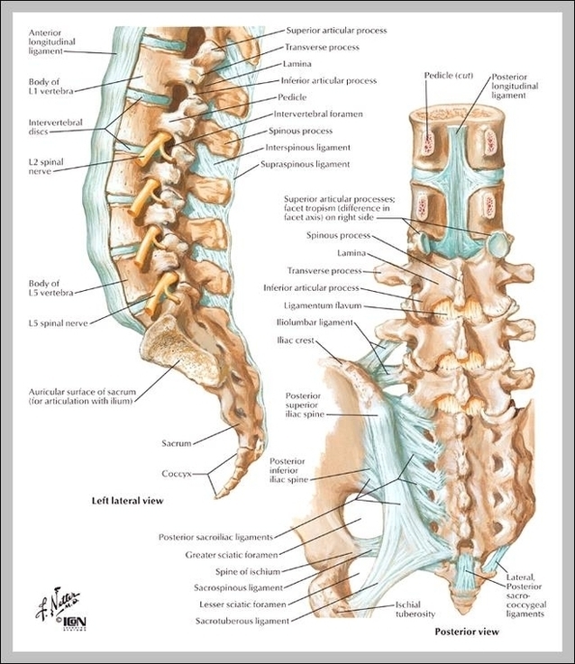
Sacral Spine Image
The strong muscles that move the ankle originate in the leg. The most well-known are the gastrocnemius and soleus muscles, which are the strong calf muscles that allow you to push up on your toes.
The anatomy of the foot is incredibly complex. This introduction to the anatomy of the foot and ankle will be very general and highlight the most relevant structures. The ankle joint or tibiotalar joint is formed where the top of the talus (the uppermost bone in the foot) and the tibia (shin bone) and fibula meet.
The anatomy of the foot is incredibly complex. This introduction to the anatomy of the foot and ankle will be very general and highlight the most relevant structures. The ankle joint or tibiotalar joint is formed where the top of the talus (the uppermost bone in the foot) and the tibia (shin bone) and fibula meet.