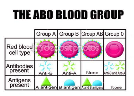
• Other gene which plays an important role in the determination of ABO blood groups is H gene with locus on chromosome 19. • Each individual inherits two ABO genes…
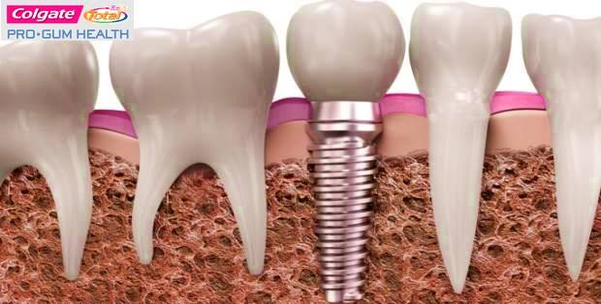
Parts of a Dental Implant: The Implant, Abutment, and Crown. While you do get a new tooth, that tooth is not actually referred to as the dental implant — the…
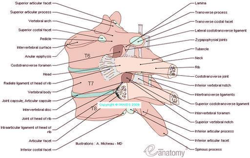
Costovertebral joint consists of the head of the rib (the head of a typical rib has two facets - each facet with a separate synovial joint separated by a ridge.…
Colonoscopy is one of the colorectal cancer screening tests available to people in the US who are over 50 years of age. Although widely touted in the US as the…
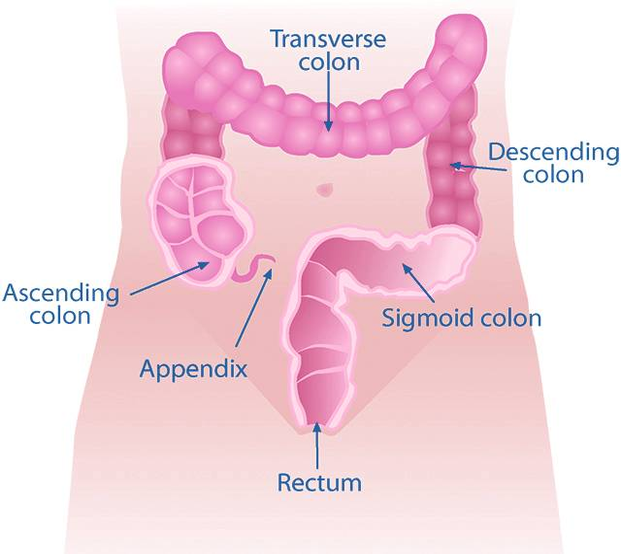
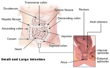
Colon: The long, coiled, tubelike organ that removes water from digested food. The remaining material, solid waste called stool, moves through the colon to the rectum and leaves the body…
Anatomy of Colon and Rectum. The rectum is the last anatomic segment before the anus. The ascending and descending colon are supported by peritoneal folds called mesentery. The right colon…
Vertebral column. The vertebral column, also known as the spinal column, is a flexible column that encloses the spinal cord and also supports the head. It consists of various groups…
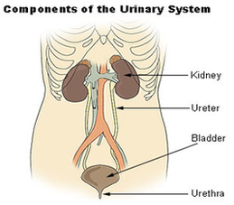
Urinary Tract Diagram: A gender-specific visual representation of the urinary tract, these types of diagrams can contain specific parts of the urinary tract such as: Kidney Diagram: An illustration of…
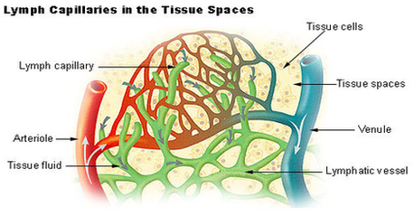
Structure of Lymphatic Capillaries. Lymph or lymphatic capillaries are tiny thin-walled vessels, closed at one end and located in the spaces between cells throughout the body. These are particularly dense…
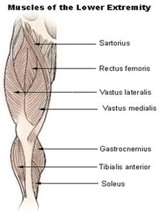
The primary muscle in this part of the body is the gastrocnemius, which gives the calf its signature bulging, muscular appearance. The anterior tibial, posterior tibial and fibular arteries are…
The specific anatomy of the large intestines is best described by referencing the individual parts, which are described in more detail below. The large intestine is roughly a meter and…
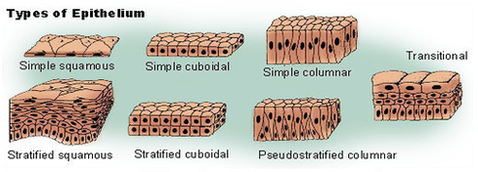
Structure of Epithelial Tissue Epithelial tissue is formed from a tightly fitted continuous layer of cells. One surface of the epithelial tissue is exposed to either the external environment or…
Some people like to diagram Scripture grammatically, like in the picture below. If that’s your thing, then by all means, go for it. You might find this book helpful (and…
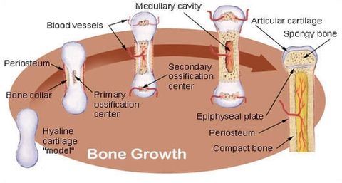
Bone Growth. Bones grow in length at the epiphyseal plate by a process that is similar to endochondral ossification. The cartilage in the region of the epiphyseal plate next to…
Venn diagrams are also called logic or set diagrams and are widely used in set theory, logic, mathematics, businesses, teaching, computer science, and statistics. Let's learn about Venn diagrams, their…
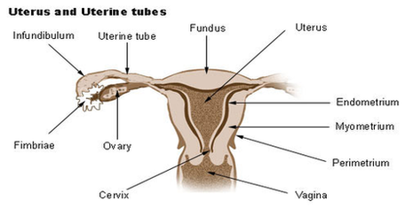
Browse 230 uterus diagram stock photos and images available or start a new search to explore more stock photos and images. An anatomical diagram depicts the method of extracting a…
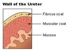
The elementary structure of the ureter is elastic muscles entangled in fiber layers, that allow to control the sphincter. The muscular layers cover the whole path between the kidney to…
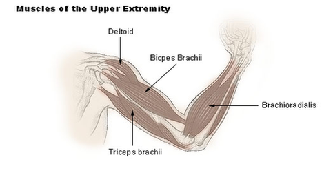
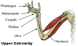
Upper limb muscles and movements. The upper limb (upper extremity) is truly a complex part of human anatomy. It is best studied broken down into its components: regions, joints, muscles,…
The forearm is the portion between the elbow and wrist. The thigh is the portion of the lower extremity between the hip and knee, and the calf is the portion…
The forearm is the portion between the elbow and wrist. The thigh is the portion of the lower extremity between the hip and knee, and the calf is the portion…