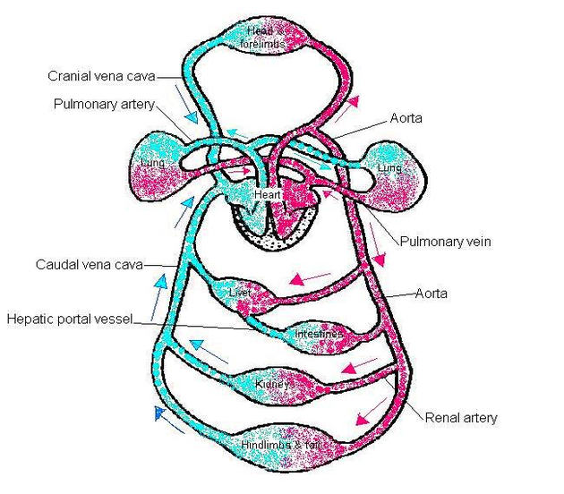
upper extremity muscles diagram

Upper extremity muscles diagram

Upper limb muscles and movements. The upper limb (upper extremity) is truly a complex part of human anatomy. It is best studied broken down into its components: regions, joints, muscles,…
upper extremity diagram1

Upper extremity diagram

The forearm is the portion between the elbow and wrist. The thigh is the portion of the lower extremity between the hip and knee, and the calf is the portion…
upper extremity diagram

Upper extremity diagram

The forearm is the portion between the elbow and wrist. The thigh is the portion of the lower extremity between the hip and knee, and the calf is the portion…
trunk muscles diagram

Trunk muscles diagram

Ventral trunk muscles (overview) The trunk (torso) is the central part of the body to which the head and the limbs are attached. Except for the brain, the trunk houses…
tonsils diagram

Tonsils diagram

sagittal view of tonsils and throat anatomy. [edit on Wikidata] Tonsils are collections of lymphoid tissue facing into the aerodigestive tract. The set of lymphatic tissue known as Waldeyer's tonsillar…
thyroid parathyroid diagram

Thyroid parathyroid diagram

The parathyroid glands are a collection of four round glands around the size of a grain of rice. These glands are attached to the posterior surface of the lateral lobes…
thymus diagram

Thymus diagram

The thymus gland is a small organ behind the breastbone that plays an important function both in the immune system and endocrine system. The growth and development of thymus continues…
thoracic cage diagram

Thoracic cage diagram

The angles of the ribs form the most posterior extent of the thoracic cage. In the anatomical position, the angles align with the medial border of the scapula. A shallow…
testis diagram

Testis diagram

The testes or testicles are part of the male genitalia or sexual reproductive organ. It is a structure that is ovoid in shape and between four to six centimeters in…
testis 2 diagram

Testis (2) diagram

The testes are covered by outer fibrous tunica vaginalis, middle tunica albuginea, and inner tunica vasculosa. Internally, the testes are divided into about 250-300 pyramid-shaped compartments called testicular lobules. On…
systemic circuit diagram

Systemic circuit diagram

The systemic circuit is that part of your circulatory system that carries blood away from your heart, delivers it to most of your organs and tissues, and returns it to…
synovial joint diagram

Synovial joint diagram

It is the most common type of joint found in the human body, and contains several structures which are not seen in fibrous or cartilaginous joints. In this article we…
stomach diagram

Stomach diagram

Given below is a labeled diagram of the stomach to help you understand stomach anatomy. The stomach is divided into four parts. These include: Cardia refers to the section of…
spleen diagram

Spleen diagram

Spleen histology slide (labeled) The spleen is a fist sized organ located in the left upper quadrant of the abdomen. It is the largest lymphoid organ and thus the largest…
reproductive male diagram

Reproductive male diagram

The male reproductive system consists of a pair of the testis, the duct system, accessory glands, and external genitalia. Let us understand in detail the male reproductive system through the…
reproductive female diagram

Reproductive female diagram

The female reproductive system is composed of a pair of ovaries along with oviducts, vagina, cervix, uterus, and the external genitalia that are located in the pelvic region. Female anatomy…
pulmonary circuit diagram

Pulmonary circuit diagram

Pulmonary circuit: Diagram of pulmonary circulation. Oxygen-rich blood is shown in red; oxygen-depleted blood in blue. Systemic circulation is the movement of blood from the heart through the body to…
planes diagram

Planes diagram

You might find a V-g diagram in your flight manual if you are flying a high performance fighter. The V-g diagram leads you to cornering speed and that allows you…
pituitary pineal glands diagram

Pituitary pineal glands diagram

Pineal and Pituitary Glands. The pineal gland is a small endocrine gland in the brain, situated beneath the back part of the corpus callosum, and secretes melatonin. The pituitary gland…
pharynx diagram

Pharynx diagram

Anatomy of the pharynx (throat). The pharynx is a hollow tube that starts behind the nose, goes down the neck, and ends at the top of the trachea and esophagus.…