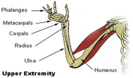
Upper Extremity Diagram

The forearm is the portion between the elbow and wrist. The thigh is the portion of the lower extremity between the hip and knee, and the calf is the portion between the knee and ankle. The normal arterial anatomy of the upper extremity is depicted graphically in Figure 13-1.
Muscles of the Upper Extremity. The pectoralis major, latissimus dorsi, deltoid, and rotator cuff muscles connect to the humerus and move the arm. The muscles that move the forearm are located along the humerus, which include the triceps brachii, biceps brachii, brachialis, and brachioradialis.
Use these carpal bones quizzes to remember carpal bones for good! Read article The lower extremity is a region of the body containing the hip, thigh, knee, leg, ankle and foot, all of which enable us to perform movements like walking, jumping and running. Start learning about these structures right away our free quiz guides below.
