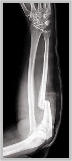
Posted inBones
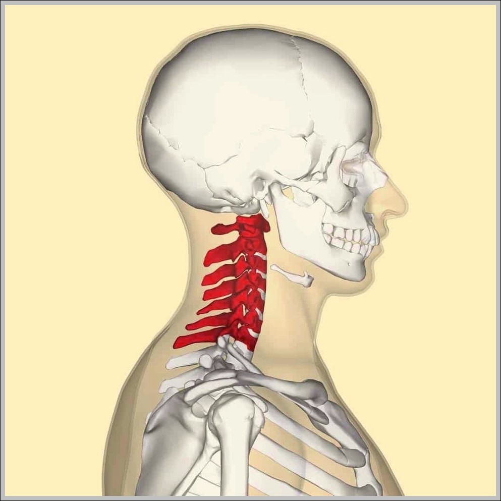
C spine picture Diagram
C-spine picture shows seven cervical vertebrae: C1 atlas (no body, anterior/posterior arches), C2 axis (dens/odontoid), C3-C6 typical (bifid spinous, transverse foramina), C7 prominent spinous (vertebra prominens).