Posted inDiagrams
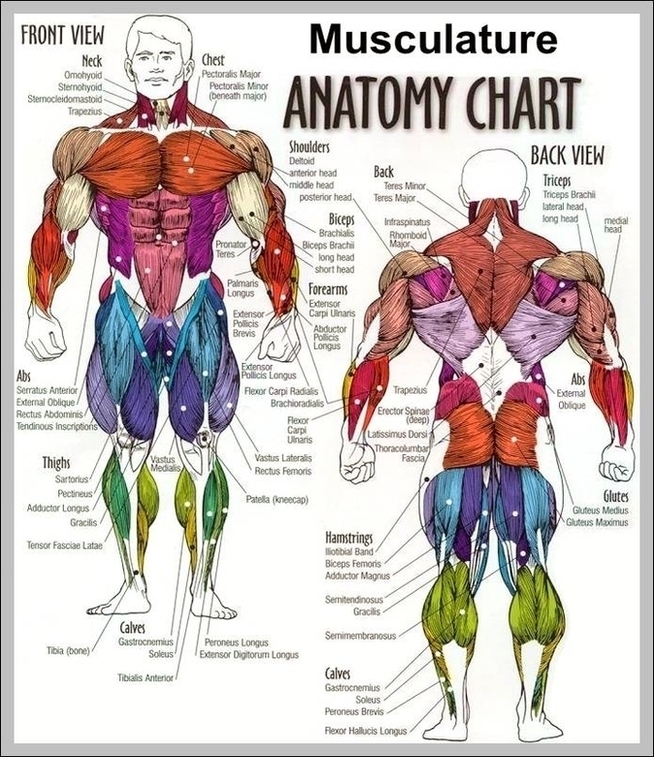
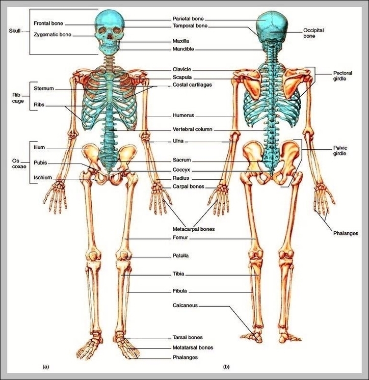
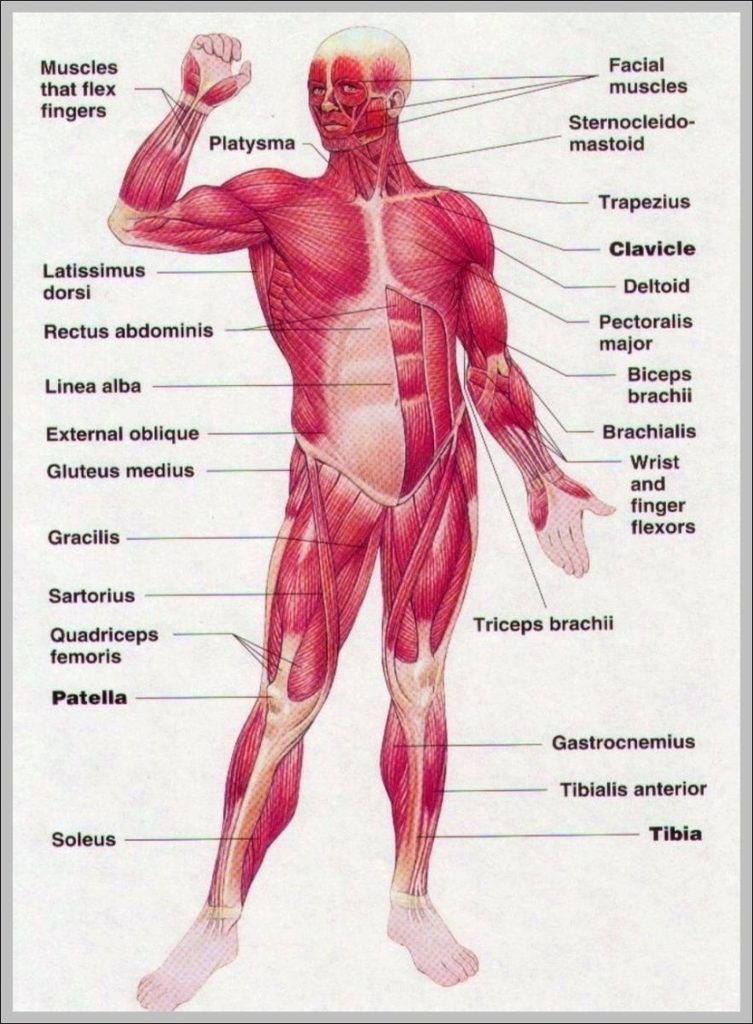
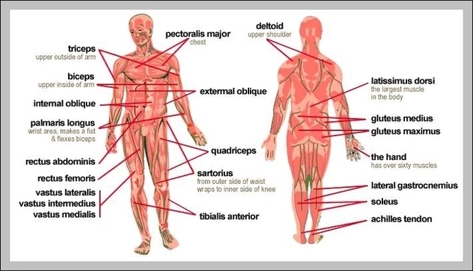
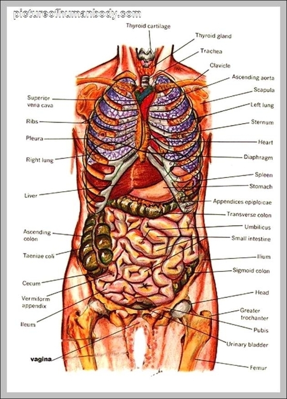
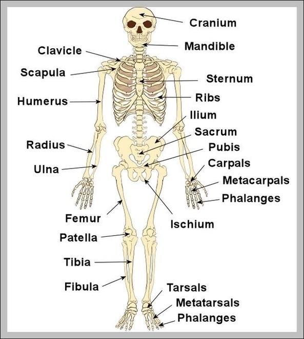
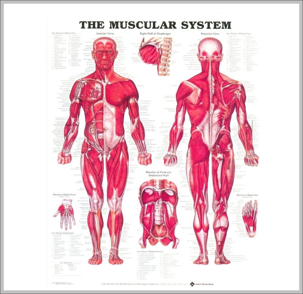
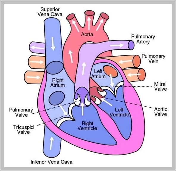
Human Body Diagram Front And Back Image
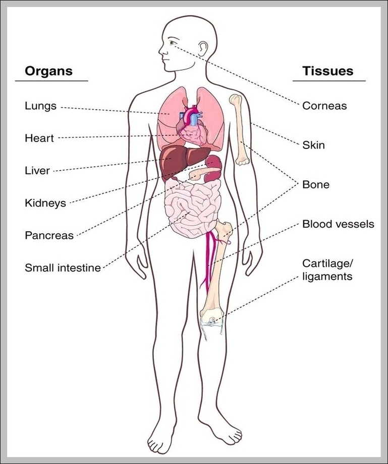
52,129 human back anatomy stock photos, vectors, and illustrations are available royalty-free. 7,751 organs of the human body diagram stock illustrations and vector graphics available royalty-free, or start a new…