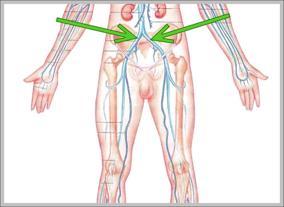
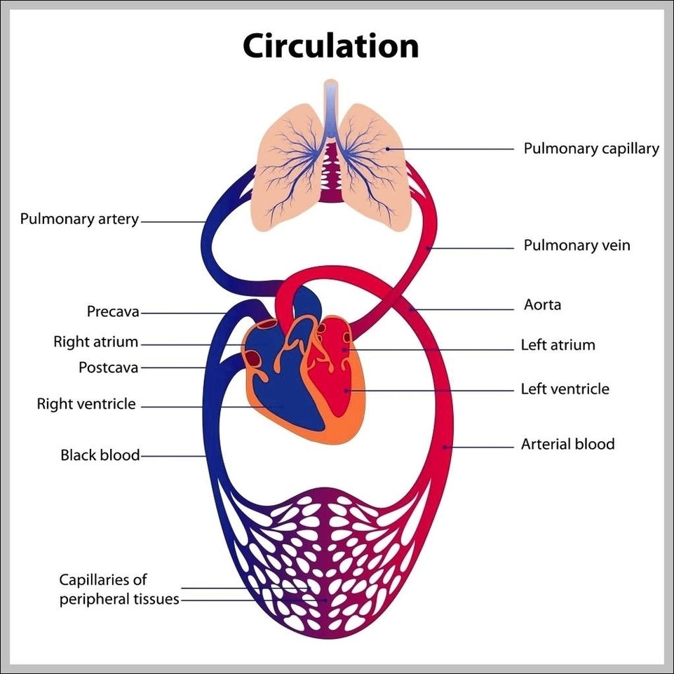
Arm Muscles Diagram Image

What Are the Muscles of Your Arms? There are four main muscles of your arms: biceps, triceps, forearm flexors, and forearm extensors. There are also a handful of other muscles that support these main four.
The human arm is divided into two main regions, the portion from the elbow to the wrist known as the forearm, and the segment from the shoulder to the elbow referred to as the arm. Arm muscle anatomy enables the arm to perform a variety of movements, including flexion, extension, pronation, and supination.
Arm muscle anatomy enables the arm to perform a variety of movements, including flexion, extension, pronation, and supination. The forearm flexors include the biceps brachii, coracobrachialis, and brachialis muscles, which help to bend the arm at the elbow, as when flexing one’s arm to show off the muscles.

Arm Muscles Diagram Image Diagram - Arm Muscles Diagram Image Chart - Human anatomy diagrams and charts explained. This anatomy system diagram depicts Arm Muscles Diagram Image with parts and labels. Best diagram to help learn about health, human body and medicine.

Arm Muscles Diagram Image

Diagram Of A Brain Image

26,763 brain diagram stock photos, vectors, and illustrations are available royalty-free.
The diagram of the brain is useful for both Class 10 and 12. It is one among the few topics having the highest weightage of marks and is frequently asked in the examinations. A well-labelled diagram of a human brain is given below for further reference.
WebMD’s Brain Anatomy Page provides a detailed diagram and definition of the brain including its function, parts, and conditions that affect it. Skip to main content Check Your Symptoms Find A Doctor Find A Dentist Connect to Care Find Lowest Drug Prices Health A-Z Health A-Z Common Conditions ADD/ADHD Allergies

Diagram Of A Brain Image Diagram - Diagram Of A Brain Image Chart - Human anatomy diagrams and charts explained. This anatomy system diagram depicts Diagram Of A Brain Image with parts and labels. Best diagram to help learn about health, human body and medicine.

Diagram Of A Brain Image

Function Of Integumentary System Image

Overall, the integumentary system functions to guard the body, providing a barrier to infection and shielding the body against temperature changes and the adverse effects of potentially harmful substances (such as UV light).
The integumentary system is composed of the following parts: 1 Skin. 2 Skin appendages. 3 Subcutaneous tissue and deep fascia. 4 Mucocutaneous junctions. 5 Breasts.
Diseases of the Integumentary System. Diseases of the integumentary system can arise from pathogenic infections, injury due to radiation, chemicals or from genetic disorders. The most common bacterial infection of the skin is probably acne.

Function Of Integumentary System Image Diagram - Function Of Integumentary System Image Chart - Human anatomy diagrams and charts explained. This anatomy system diagram depicts Function Of Integumentary System Image with parts and labels. Best diagram to help learn about health, human body and medicine.

Function Of Integumentary System Image

Body Bone Structure Image

Browse 67,060 human bone stock photos and images available, or search for human bone marrow or human bone structure to find more great stock photos and pictures.
Bone Structure. 1 Gross Anatomy of Bone. The structure of a long bone allows for the best visualization of all of the parts of a bone ( (Figure) ). A long bone has two … 2 Bone Markings. 3 Bone Cells and Tissue. 4 Compact and Spongy Bone.
Microscopic Structure of Bones. The skeleton makes up about 30-40% of an adult’s body mass. The skeleton’s mass is made up of nonliving bone matrix and many tiny bone cells. Roughly half of the bone matrix’s mass is water, while the other half is collagen protein and solid crystals of calcium carbonate and calcium phosphate.

Body Bone Structure Image Diagram - Body Bone Structure Image Chart - Human anatomy diagrams and charts explained. This anatomy system diagram depicts Body Bone Structure Image with parts and labels. Best diagram to help learn about health, human body and medicine.

Body Bone Structure Image

Body Chart Organs Image

Browse 12,251 human body organs anatomy stock photos and images available, or start a new search to explore more stock photos and images.
Human internal organs vector Vector isolated illustration of human internal organs in male body. Stomach, liver, intestine, bladder, lung, testicle, uterus, spine, pancreas, kidney, heart, bladder icon. Donor medical poster organs of the human body diagram stock illustrations
Vital organs 1 Brain. The brain is the body’s control center. … 2 Heart. The heart is the most important organ of the circulatory system, which helps deliver blood to the body. 3 Lungs. The lungs work with the heart to oxygenate blood. … 4 Liver. The liver is the most important organ of the metabolic system. … 5 Kidneys. …

Body Chart Organs Image Diagram - Body Chart Organs Image Chart - Human anatomy diagrams and charts explained. This anatomy system diagram depicts Body Chart Organs Image with parts and labels. Best diagram to help learn about health, human body and medicine.

Body Chart Organs Image

Anatomy Lessons Image

Anatomy Lessons Image Diagram - Anatomy Lessons Image Chart - Human anatomy diagrams and charts explained. This anatomy system diagram depicts Anatomy Lessons Image with parts and labels. Best diagram to help learn about health, human body and medicine.

Anatomy Lessons Image

Brachial Fossa Image

The antecubital fossa is bounded on the top by the skin and several veins and nerves, while it is bounded on the bottom by more muscles. The antecubital fossa houses several important structures. The radial nerve passes on the same side of the arm as the radius. The radial nerve supplies many of the muscles of the arm.
It arises from the distal part of the bone, below your biceps brachii muscle. It then courses down the front of your arm, over your elbow joint, and inserts on the coronoid process and tuberosity of your ulna. The brachialis muscle, along with the supinator muscle, makes up the floor of the cubital fossa of your elbow.
The brachialis is a muscle in the front of your elbow that flexes, or bends, the joint. It does this when your forearm is in a palm down, pronated, position.

Brachial Fossa Image Diagram - Brachial Fossa Image Chart - Human anatomy diagrams and charts explained. This anatomy system diagram depicts Brachial Fossa Image with parts and labels. Best diagram to help learn about health, human body and medicine.

Brachial Fossa Image

Pictures Of Female Anatomy Image

Pictures Of Female Anatomy Image Diagram - Pictures Of Female Anatomy Image Chart - Human anatomy diagrams and charts explained. This anatomy system diagram depicts Pictures Of Female Anatomy Image with parts and labels. Best diagram to help learn about health, human body and medicine.

Pictures Of Female Anatomy Image

Study Human Anatomy Image

Human anatomy is the scientific study of human body structures. The two subdivisions of human anatomy are gross anatomy and microscopic anatomy. Gross anatomy refers to the study of human organs that can be seen without magnification. Microscopic anatomy refers to the study of small anatomic parts that can only be seen with a microscope.
Anatomy is the study of the structure of an object. Human anatomy deals with the way the parts of humans interact to form a functional unit. What do you understand by human physiology? Human Physiology deals with the mechanical, biochemical and physical functions of humans.
Browse 28,679 anatomy of the human body stock photos and images available, or start a new search to explore more stock photos and images.

Study Human Anatomy Image Diagram - Study Human Anatomy Image Chart - Human anatomy diagrams and charts explained. This anatomy system diagram depicts Study Human Anatomy Image with parts and labels. Best diagram to help learn about health, human body and medicine.

Study Human Anatomy Image

Diagram Of The Knee Muscles Image

To understand one of the most complex joints of our body i.e. the knee joint, you need a perfectly labeled diagram of the knee. This will help you to understand the mechanism as well as the working.
Knee joint is one of the most important hinge joints of our body. Its complexity and its efficiency is the best example of God’s creation. The anatomy of the knee consists of bones, muscles, nerves, cartilages, tendons and ligaments. All these parts combine and work together.
The hamstrings and quadriceps work together, one contracting (agonist) while the other relaxes (antagonist) to allow the knee to bend and straighten. Here we look at each of the muscles of the knee, how they work, what can go wrong and how to prevent knee muscle injuries.

Diagram Of The Knee Muscles Image Diagram - Diagram Of The Knee Muscles Image Chart - Human anatomy diagrams and charts explained. This anatomy system diagram depicts Diagram Of The Knee Muscles Image with parts and labels. Best diagram to help learn about health, human body and medicine.

Diagram Of The Knee Muscles Image

Shoulder Joint Name Image

The shoulder joint is formed by the articulation of the head of the humerus with the glenoid cavity (or fossa) of the scapula. This gives rise to the alternate name for the shoulder joint – the glenohumeral joint.
The shoulder is one of the largest and most complex joints in the body. The shoulder joint is formed where the humerus (upper arm bone) fits into the scapula (shoulder blade), like a ball and socket. Other important bones in the shoulder include:
The Shoulder Capsule. The shoulder capsule surrounds the ball-and-socket part of the shoulder joint. The capsule separates the joint from the rest of the body and contains the joint fluid. Several ligaments make up parts of the joint capsule, and these ligaments are important in keeping the shoulder joint in proper position.

Shoulder Joint Name Image Diagram - Shoulder Joint Name Image Chart - Human anatomy diagrams and charts explained. This anatomy system diagram depicts Shoulder Joint Name Image with parts and labels. Best diagram to help learn about health, human body and medicine.

Shoulder Joint Name Image

Body Anatomy Chart Organs Image

Browse 7,751 organs of the human body diagram stock illustrations and vector graphics available royalty-free, or start a new search to explore more great stock images and vector art. internal organs and circulatory system Vector isolated illustration of human internal organs and circulatory system in man body.
Human internal organs vector Vector isolated illustration of human internal organs in male body. Stomach, liver, intestine, bladder, lung, testicle, uterus, spine, pancreas, kidney, heart, bladder icon. Donor medical poster organs of the human body diagram stock illustrations
Browse 5,814 male human anatomy diagram stock photos and images available, or start a new search to explore more stock photos and images. Male silhouette (contour) on white background, vector. Male, female and children’s silhouette on white background,…

Body Anatomy Chart Organs Image Diagram - Body Anatomy Chart Organs Image Chart - Human anatomy diagrams and charts explained. This anatomy system diagram depicts Body Anatomy Chart Organs Image with parts and labels. Best diagram to help learn about health, human body and medicine.

Body Anatomy Chart Organs Image

Types Of Muscles Image

Types Of Muscles Image Diagram - Types Of Muscles Image Chart - Human anatomy diagrams and charts explained. This anatomy system diagram depicts Types Of Muscles Image with parts and labels. Best diagram to help learn about health, human body and medicine.

Types Of Muscles Image

Diagram Of Skeleton Image

Browse 2,854 human skeleton diagram stock photos and images available, or start a new search to explore more stock photos and images. human anatomy skeleton and muscles of the body – human skeleton diagram stock illustrations
Skeletal System Diagram. Skeletal system diagrams are illustrations of the human skeleton, used mostly for educational purposes or in presentations. Skeletal diagrams are tools used by students to learn and study all 206 bones (this number can vary from person to person) of the human body.
Lateral View Skeletal Diagram: A lateral view skeletal diagram offers a side view of the human skeleton. Skeletal diagrams can also be used to show bone development or growth which begins en utero.

Diagram Of Skeleton Image Diagram - Diagram Of Skeleton Image Chart - Human anatomy diagrams and charts explained. This anatomy system diagram depicts Diagram Of Skeleton Image with parts and labels. Best diagram to help learn about health, human body and medicine.

Diagram Of Skeleton Image

Back Muscles Diagram Image

25,649 back muscle anatomy stock photos, vectors, and illustrations are available royalty-free.
Certain back muscles extend to other areas, like the shoulders, upper arms, and thighs. There are three different types of muscles in the body: the heart muscle, smooth muscles, and skeletal muscles. The back muscles are skeletal muscles. They support bones, in this case, the vertebrae.
It comprises the vertebral column (spine) and two compartments of back muscles; extrinsic and intrinsic. The back functions are many, such as to house and protect the spinal cord, hold the body and head upright, and adjust the movements of the upper and lower limbs .

Back Muscles Diagram Image Diagram - Back Muscles Diagram Image Chart - Human anatomy diagrams and charts explained. This anatomy system diagram depicts Back Muscles Diagram Image with parts and labels. Best diagram to help learn about health, human body and medicine.

Back Muscles Diagram Image

Migraines And Headaches Diagram

Migraines And Headaches Diagram Diagram - Migraines And Headaches Diagram Chart - Human anatomy diagrams and charts explained. This anatomy system diagram depicts Migraines And Headaches Diagram with parts and labels. Best diagram to help learn about health, human body and medicine.

Migraines and Headaches Diagram

Women Reproductive Organs Anatomy

Women Reproductive Organs Anatomy Diagram - Women Reproductive Organs Anatomy Chart - Human anatomy diagrams and charts explained. This anatomy system diagram depicts Women Reproductive Organs Anatomy with parts and labels. Best diagram to help learn about health, human body and medicine.

Women reproductive organs anatomy

Vagina Anatomy Diagram

Vagina Anatomy Diagram Diagram - Vagina Anatomy Diagram Chart - Human anatomy diagrams and charts explained. This anatomy system diagram depicts Vagina Anatomy Diagram with parts and labels. Best diagram to help learn about health, human body and medicine.

Vagina anatomy diagram

Bronchus Anatomy Diagram With Labels

Bronchus Anatomy Diagram With Labels Diagram - Bronchus Anatomy Diagram With Labels Chart - Human anatomy diagrams and charts explained. This anatomy system diagram depicts Bronchus Anatomy Diagram With Labels with parts and labels. Best diagram to help learn about health, human body and medicine.

Bronchus Anatomy Diagram with labels

Human Skull Diagram

Human Skull Diagram Diagram - Human Skull Diagram Chart - Human anatomy diagrams and charts explained. This anatomy system diagram depicts Human Skull Diagram with parts and labels. Best diagram to help learn about health, human body and medicine.

Human Skull diagram