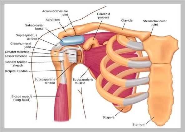
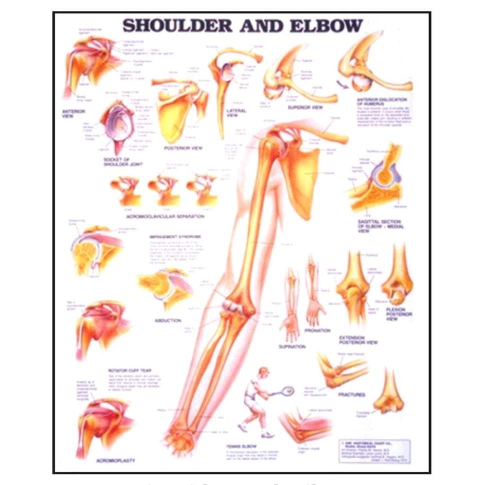
The shoulder is one of the largest and most complex joints in the body. The shoulder joint is formed where the humerus (upper arm bone) fits into the scapula (shoulder…
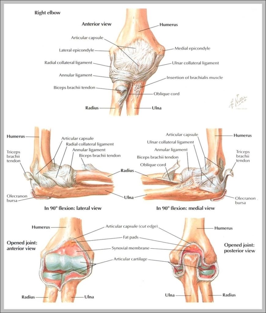
Ulnar collateral ligament or UCL, lateral collateral ligament and annular ligament form the ligaments in elbow. Here we will look in detail about the ligaments, the common injuries affecting them,…
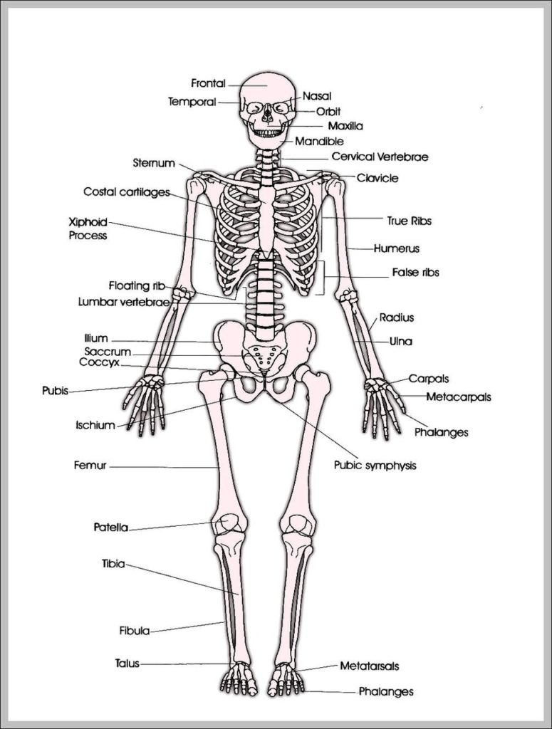
2,854 human skeleton diagram stock photos and images available, or start a new search to explore more stock photos and images. human anatomy skeleton and muscles of the body -…
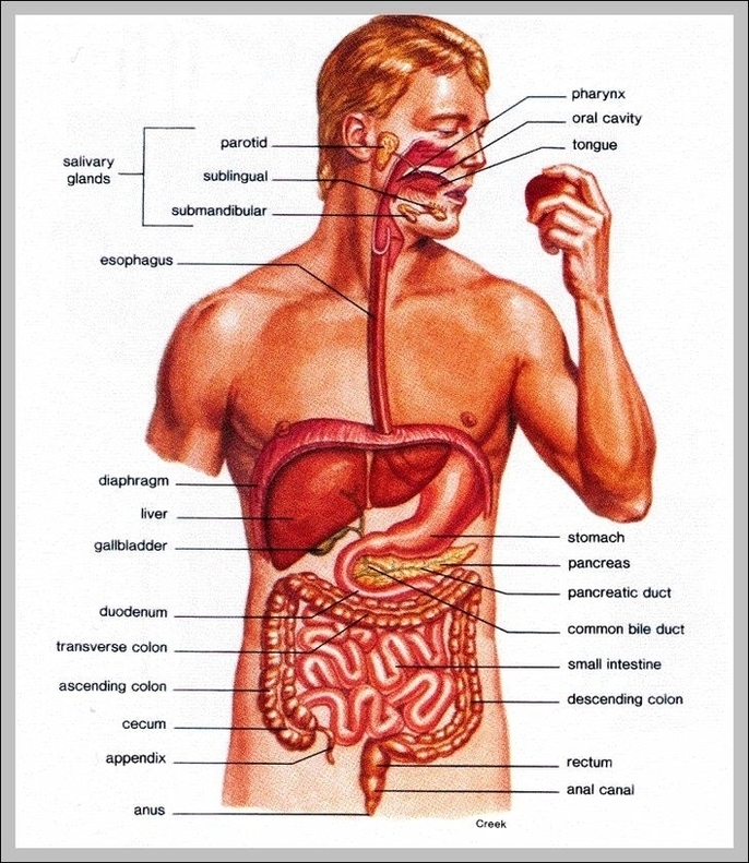
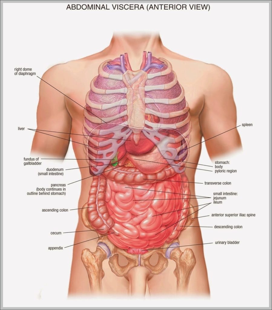
7,751 organs of the human body diagram stock illustrations and vector graphics available royalty-free, or start a new search to explore more great stock images and vector art. internal organs…
Your lower back (lumbar spine) is the anatomic region between your lowest rib and the upper part of the buttock. 1 Your spine in this region has a natural inward…
Body image refers to a person’s emotional attitudes, beliefs, and perceptions of their own body. Experts describe it as a complex emotional experience. A person’s body image will range from…
Anatomy of the Stomach. The stomach is an organ of the digestive system. It is an expanded section of the digestive tube between the esophagus and small intestine. Its characteristic…
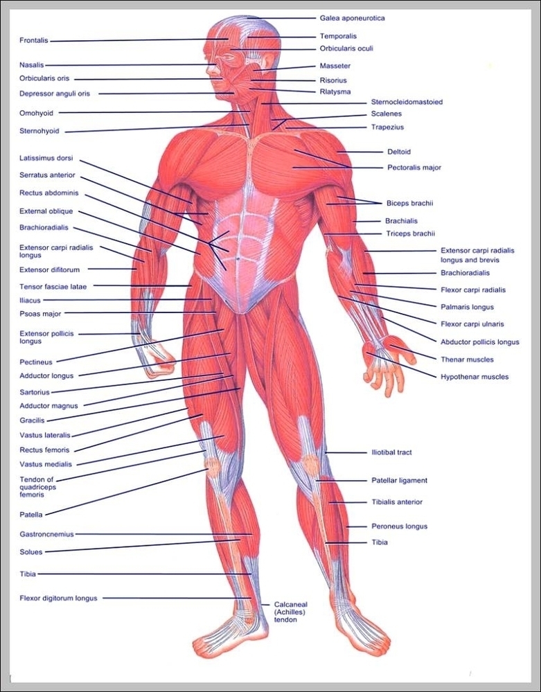
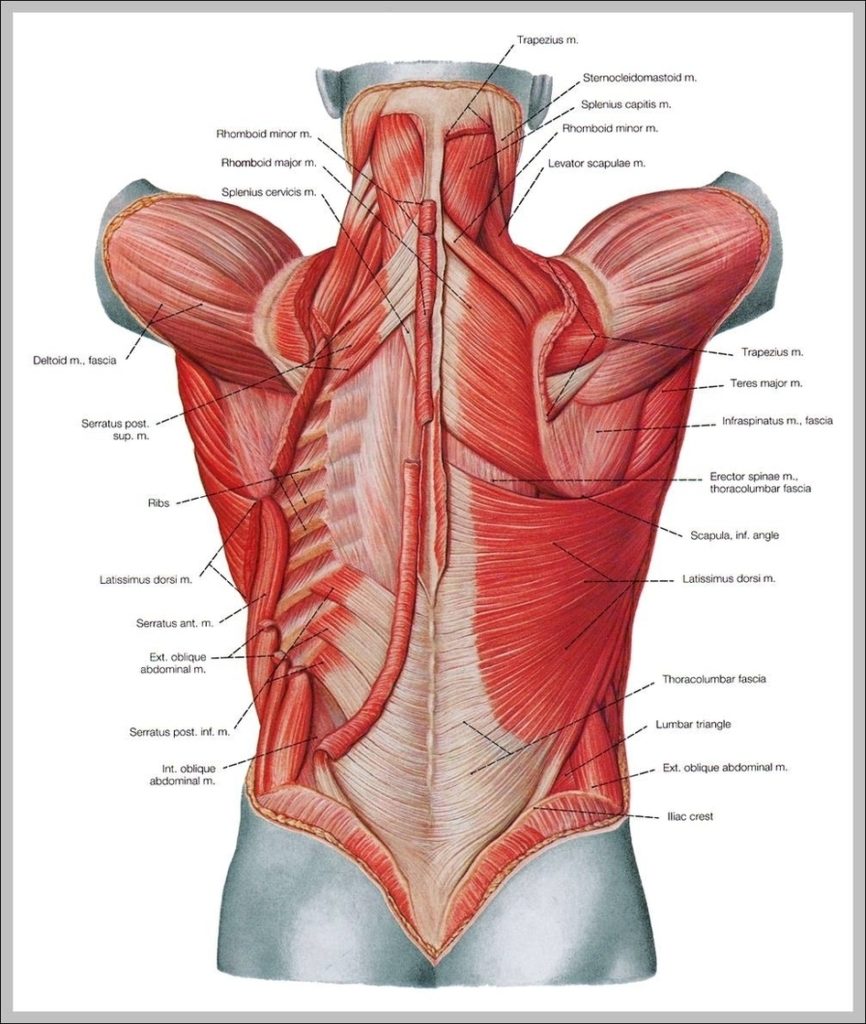
128,412 muscle anatomy stock photos, vectors, and illustrations are available royalty-free. Forearms - Anatomy Muscles Rhomboid minor and rhomboid major, levator scapulae and latissimus dorsi muscles - didactic board of…
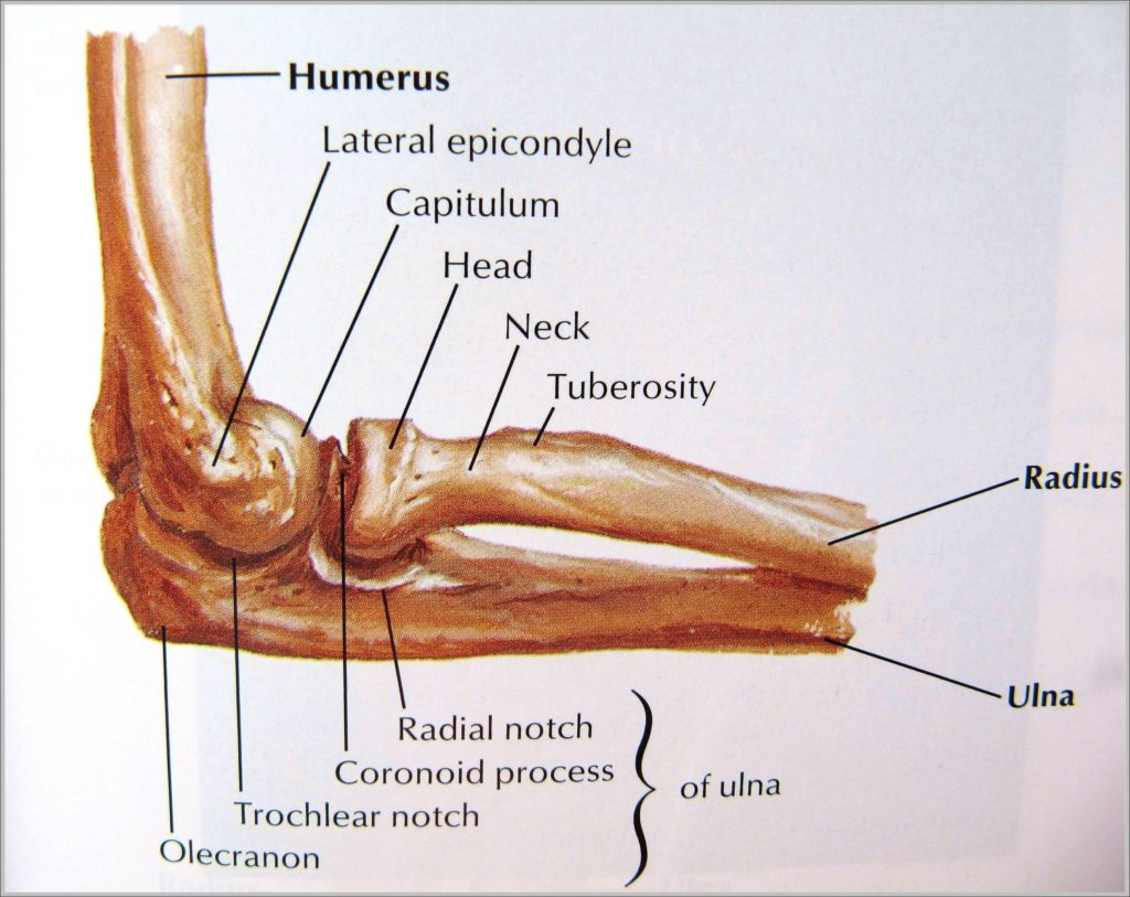
The Anatomy of the Elbow The elbow is a hinged joint made up of three bones, the humerus, ulna, and radius. The ends of the bones are covered with cartilage.…
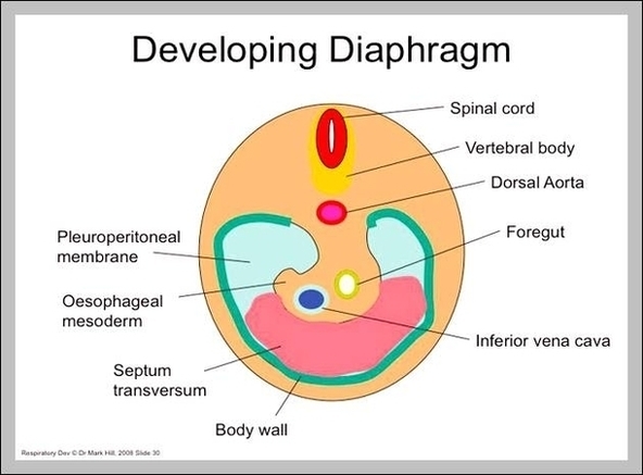
The diaphragm in the respiratory system is the dome-shaped sheet of muscle that separates the chest from the abdomen. It is also referred to the thoracic diaphragm because it’s located…
The Anatomy of the Elbow The elbow is a hinged joint made up of three bones, the humerus, ulna, and radius. The ends of the bones are covered with cartilage.…
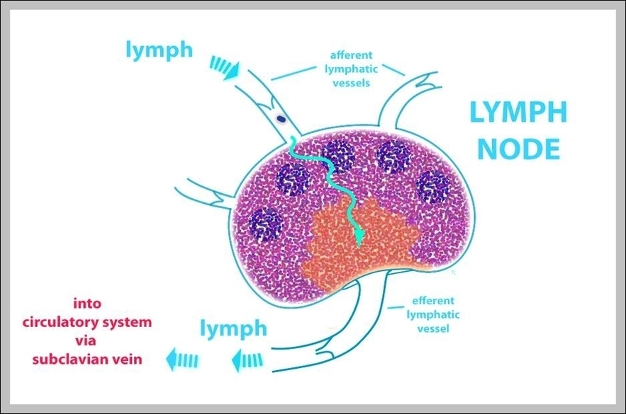
What is lymph? Lymph represents a clear, sometimes milky colored, substance that flows within the lymphatic system. Part of the lymph flowing through the lymphatic system comes from the plasma…
25,649 back muscle anatomy stock photos, vectors, and illustrations are available royalty-free. As with other parts of the body, the back has several layers of muscles. Some are closer to…
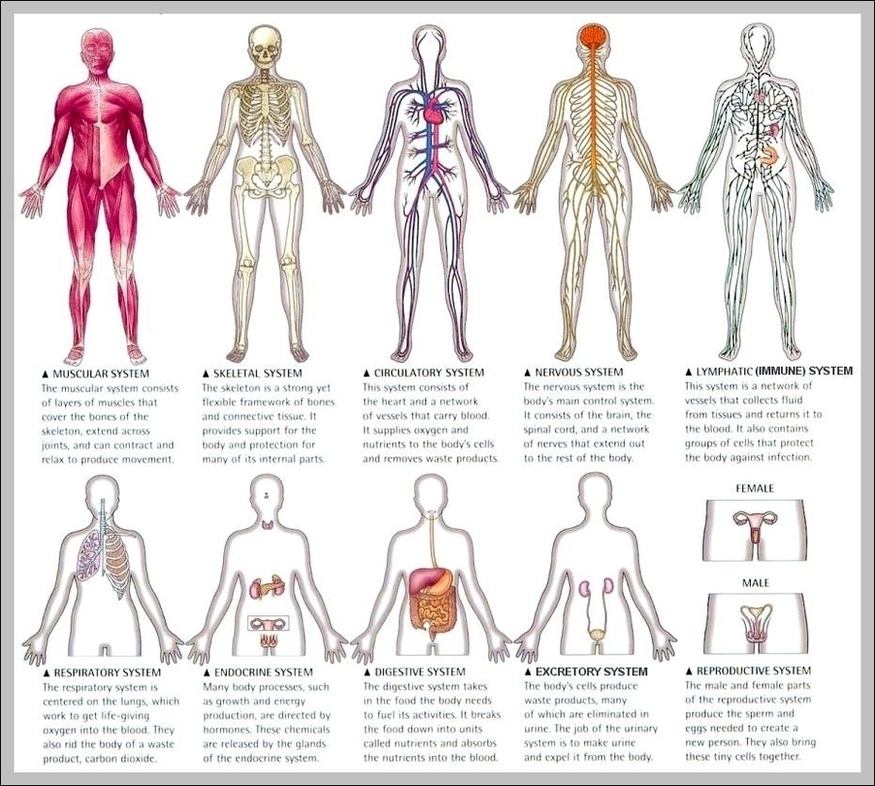
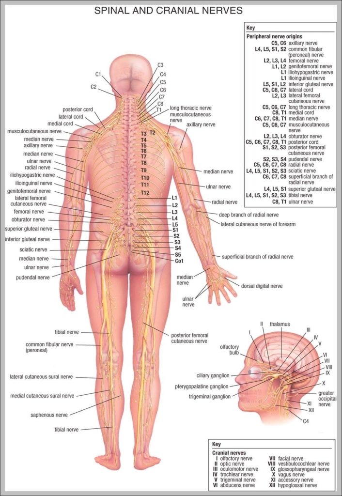
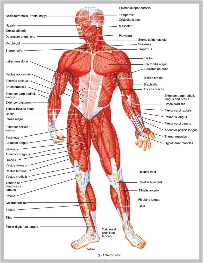
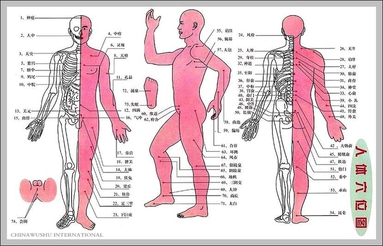
The human body comprises 12 distinct human body systems, and their functions reflect their names: cardiovascular, digestive, endocrine, immune, integumentary, lymphatic, muscular, nervous, reproductive, respiratory, skeletal and urinary. 183,298 human…
The sagital view of the brain reflects some of the inverted c-shaped rings including the cortex, cingulate gyrus, indusium griseum, corpus callosum, septum pellucidum/lateral ventricles, and the thalamus which is…
Ligaments. There are many ligament of the hand that are made up of tough bands of fibrous tissue. As there are many small bones and joints in the hand, there…
Faculty members within the Medical Laboratory Technician program are experienced in hospitals and clinical laboratories. Five - Six weeks of clinical placement will provide you with experience in a real…
How Many Muscles In the Human Body? How Many Muscles In the Human Body? There are about 700 named skeletal muscles in the human body, including roughly 400 that no…