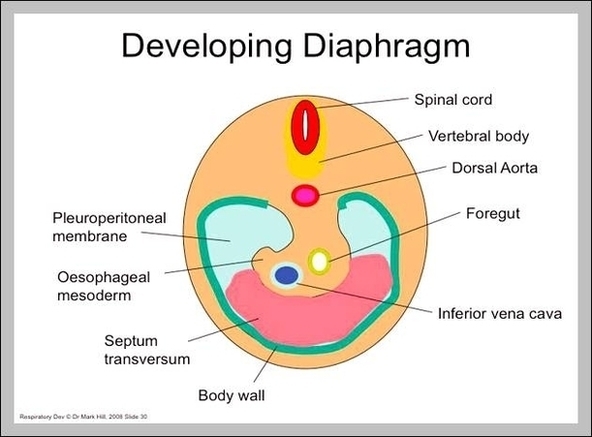
The diaphragm in the respiratory system is the dome-shaped sheet of muscle that separates the chest from the abdomen. It is also referred to the thoracic diaphragm because it’s located in the thoracic cavity, or chest. It is attached to the spine, ribs and sternum and is the main muscle of respiration,…
The thoracic diaphragm is a large, flat muscle that plays a vital role in the respiratory system, and is located just beneath the two lungs, dividing the chest cavity from the abdominal cavity. With its characteristic dome shape, it is the primary respiratory muscle, also supporting the lungs and heart.
The thoracic diaphragm is a large, flat muscle that plays a vital role in the respiratory system, and is located just beneath the two lungs, dividing the chest cavity from the abdominal cavity. With its characteristic dome shape, it is the primary respiratory muscle, also supporting the lungs and heart.
Respiratory Diaphragm Image
Posted inDiagrams
Respiratory Diaphragm Image
Post navigation
Previous Post
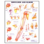
 Elbow Bone Anatomy Image
Next Post
Shoulder and Elbow Diagram