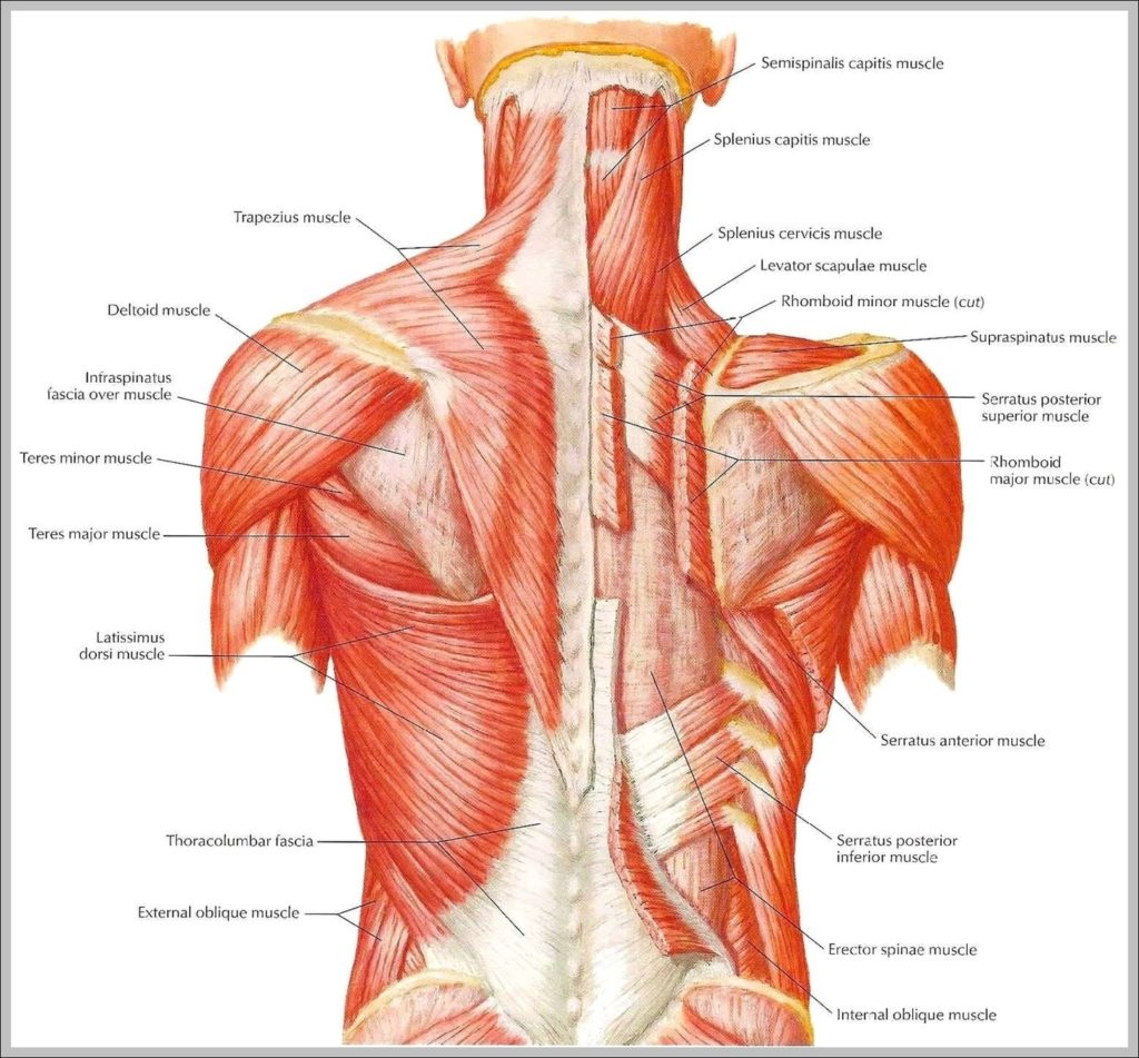
Your back consists of a complex array of bones, discs, nerves, joints, and muscles. The muscles of your back support your spine, attach your pelvis and shoulders to your trunk,…
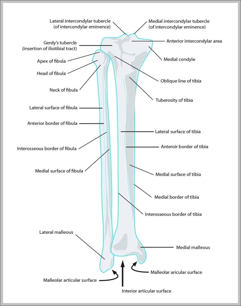
Human lower leg muscles (soleus), illustration. The bones of the the lower leg and foot. Shown are the tibia; femur; patella; fibula, medial malleolus, lateral malleolus; metatarsals; bones; lower l…
How to Digitize Plot and Graph Images? Step 1: Scanning the plot or graph to create the image. For a physical document, you have to scan the page on which…
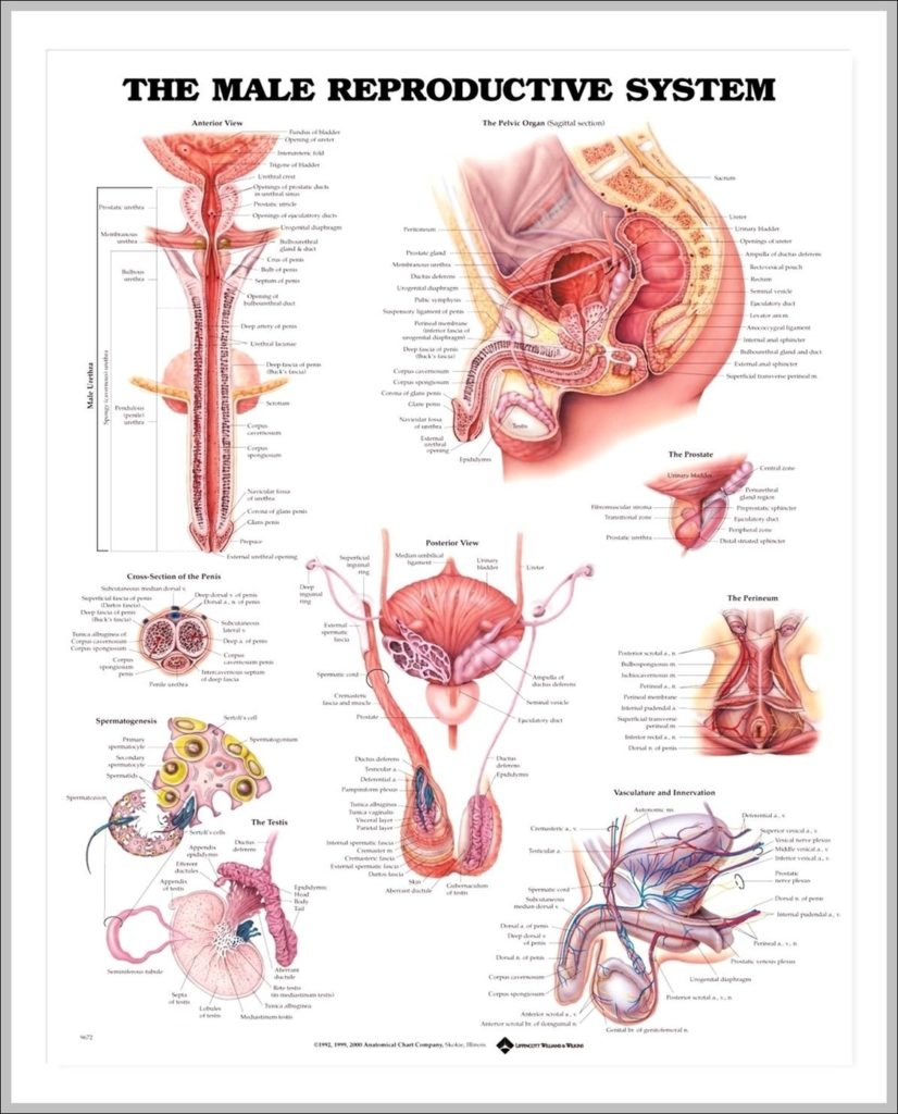
3D computer graphic cross section of male reproductive system with labels. Male reproductive system The human male reproductive system consists of a number of sex organs that form a part…
33,602 muscular system stock photos, vectors, and illustrations are available royalty-free. See muscular system stock video clips Last Updated: Jul 16, 2019 The muscular system is responsible for the movement…
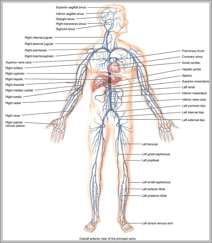
4,089 veins human body stock photos and images available, or start a new search to explore more stock photos and images. In the human body, all the veins have one-way…
Lower back muscle anatomy includes the Multifidus, Longissimus, Spinalis, and Quadratus Lumborum. The muscles of the low back work together with the transverse abdominal muscles to increase intra-abdominal pressure. The…
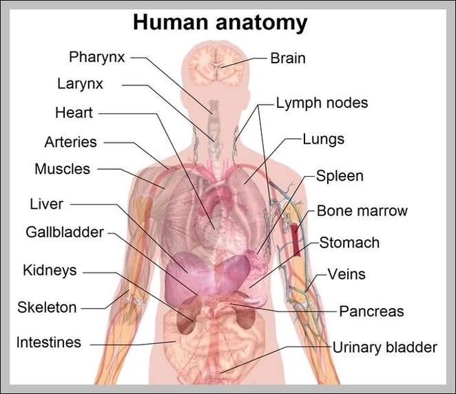
7,751 organs of the human body diagram stock illustrations and vector graphics available royalty-free, or start a new search to explore more great stock images and vector art. internal organs…
The pancreas is an organ located in the abdomen. It plays an essential role in converting the food we eat into fuel for the body's cells. The pancreas has two…
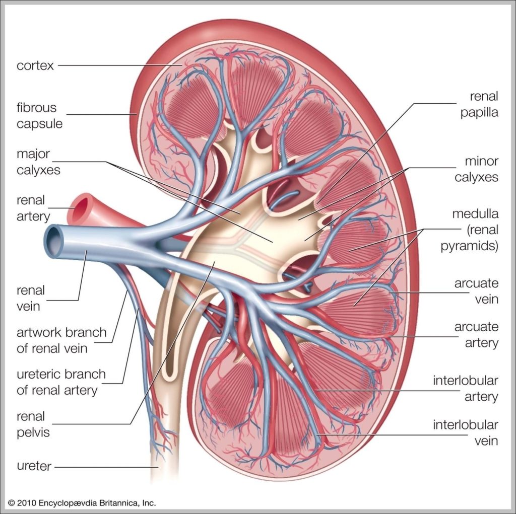
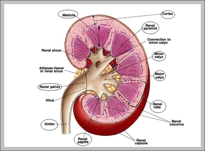
What is a Urinary System Diagram? Urinary system diagrams are illustrations of the urinary system, also referred to as the renal system. The urinary system, at a high level, contains…
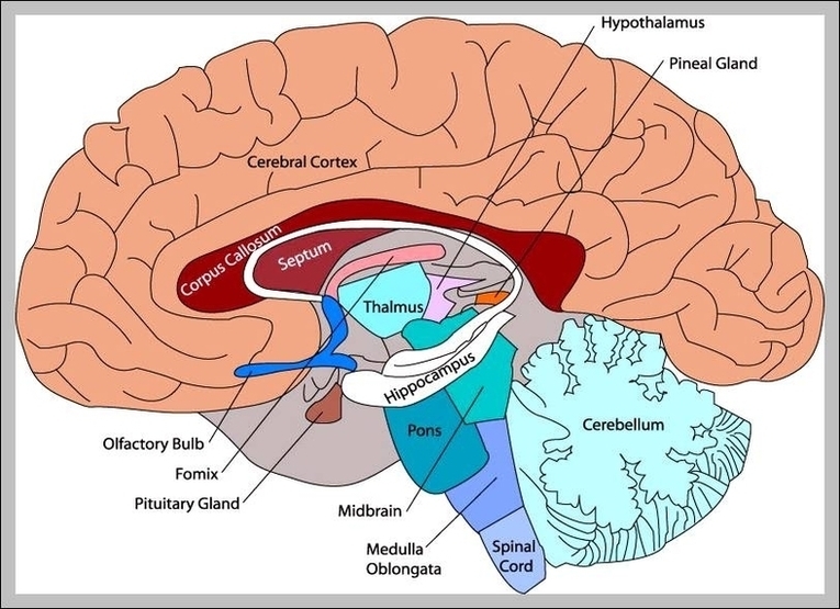
Before we have a look at the brain diagram, it is important to go through a few facts about the brain and its function. This will help you understand the…
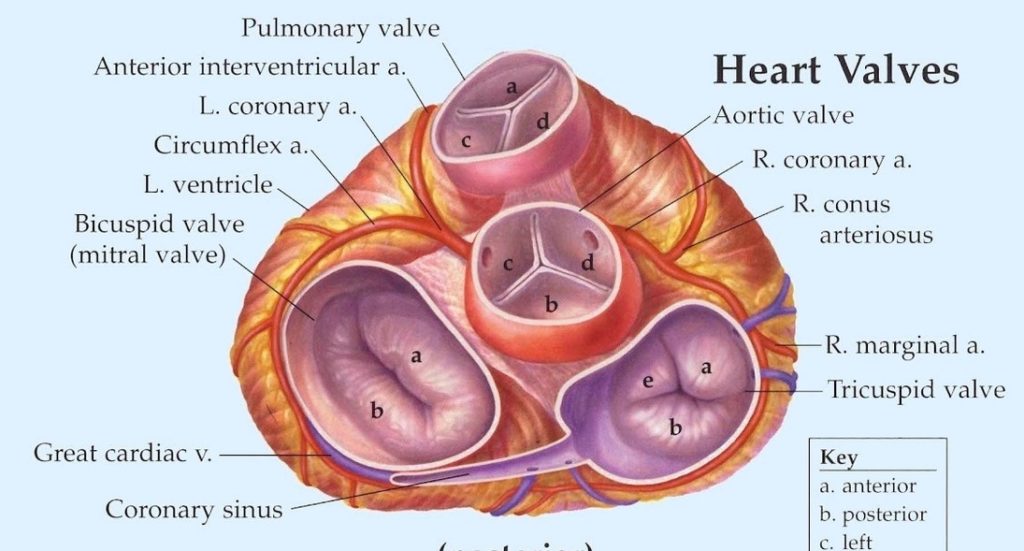
Heart Valves. The valves keep blood moving through the heart in the right direction. The mitral valve and tricuspid valve are located between the between the atria (upper heart chambers)…
All the images are in vector format, allowing an optimal web display with zoom and shifting of the anatomical images. A general view of the spine with the various levels…
12,239 human body anatomy organs stock photos and images available or start a new search to explore more stock photos and images. Lungs, heart, liver, stomach, tooth human body organ…
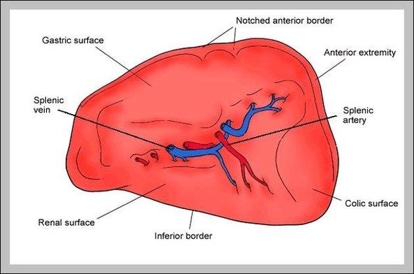
Your spleen is a small organ that sits inside your left rib cage, just above your stomach. In adults, the spleen is about the size of an avocado. The spleen…
Liver Location : Where is your liver. Consider a human body. When we remove the limbs, head and neck, what remains is our trunk. The trunk is divided into two…
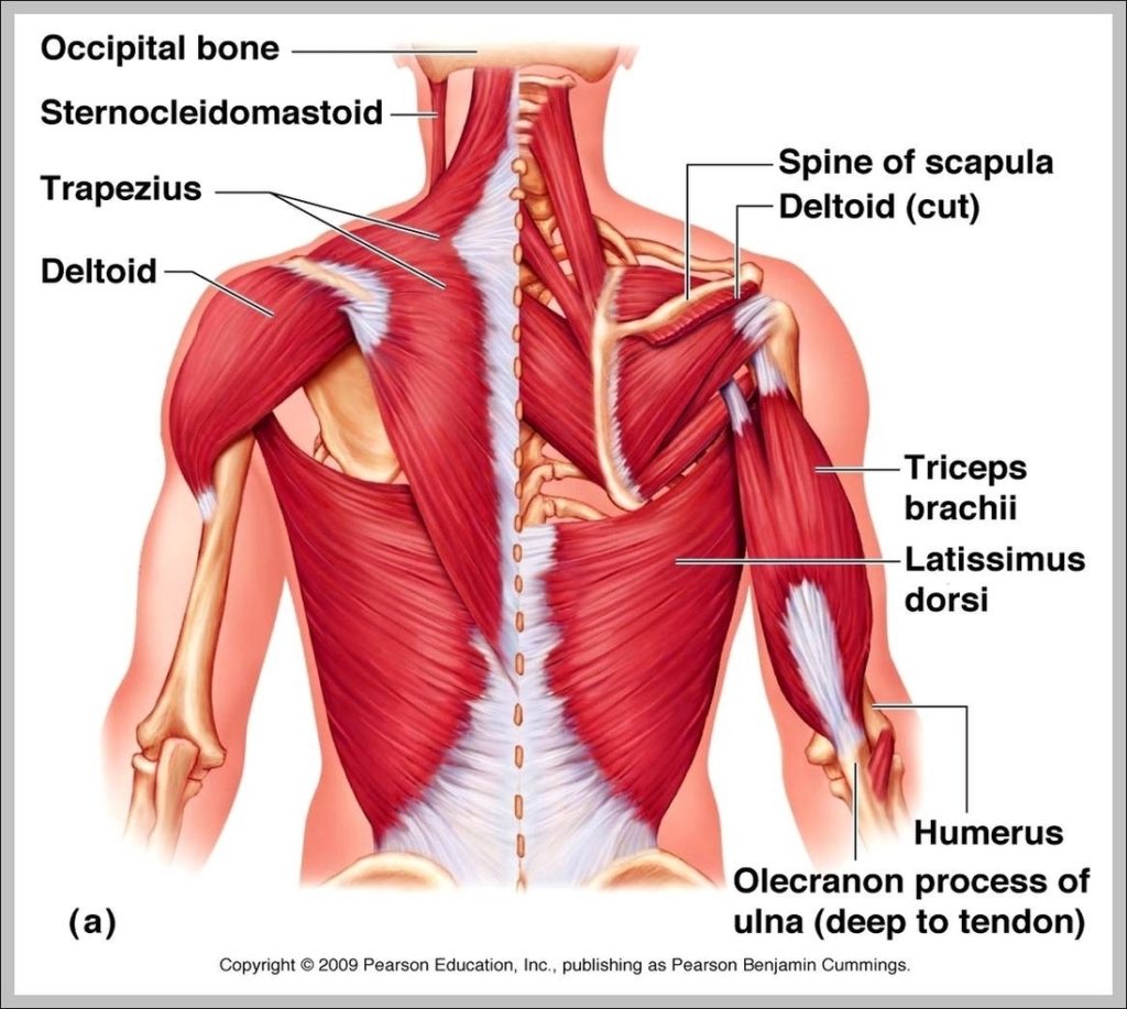
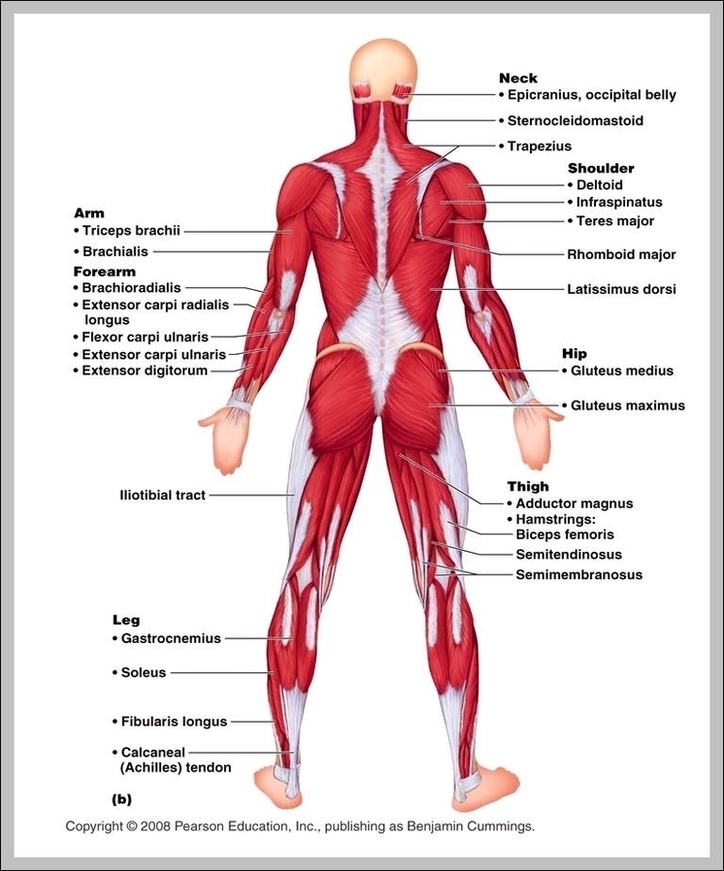
Upper Back Muscles. The deltoid, teres major, teres minor, infraspinatus, supraspinatus (not shown) and subscapularis muscles (not shown) all extend from the scapula to the humerus and act on the…
12,251 human body organs anatomy stock photos and images available, or start a new search to explore more stock photos and images. Stomach, liver, intestine, bladder, lung, testicle, spine, pancreas,…