62 labeled diagram of the skeletal system pic stock photos and images available, or start a new search to explore more stock photos and images. Skeletal Poster Human skeletal system…
2,854 human skeleton diagram stock photos and images available, or start a new search to explore more stock photos and images. human anatomy skeleton and muscles of the body -…
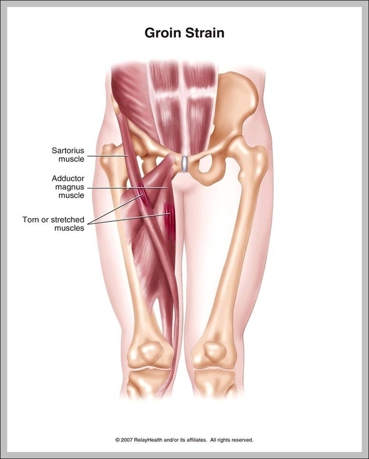
Groin muscles help support the hip joint. The groin muscles are a group of muscles situated high on the leg in the inner thigh. This group includes the adductor magnus,…
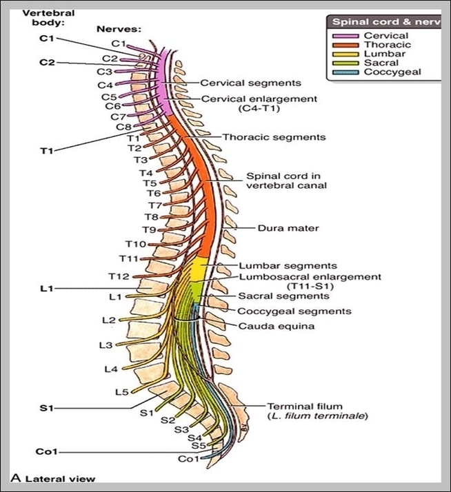
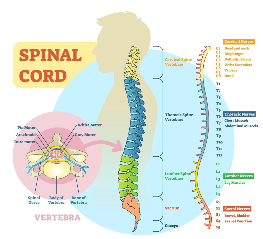
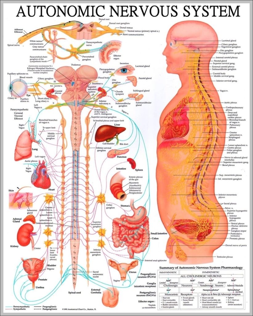
Spinal Cord. The spinal cord is a part of the central nervous system. It is a long pipe-like structure arising from the medulla oblongata, part of the brain consisting of…
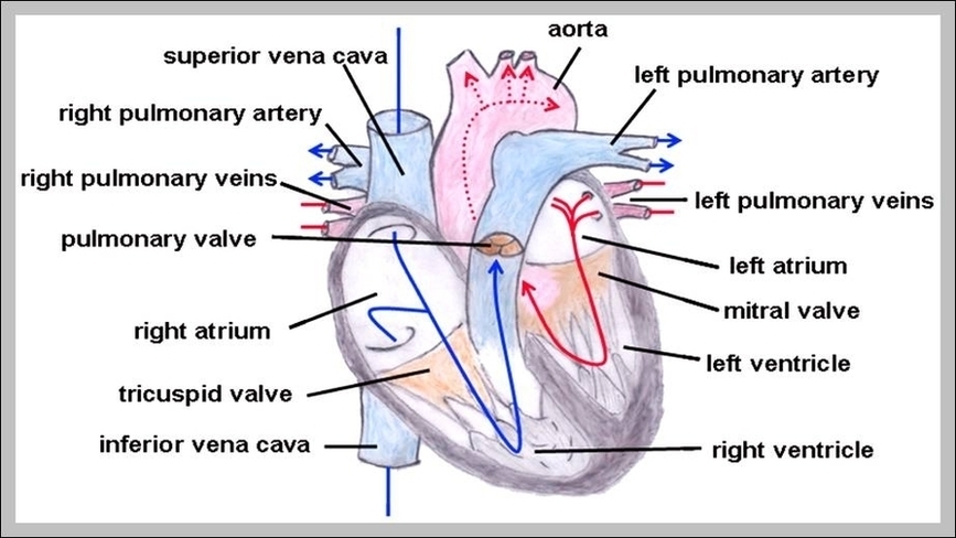
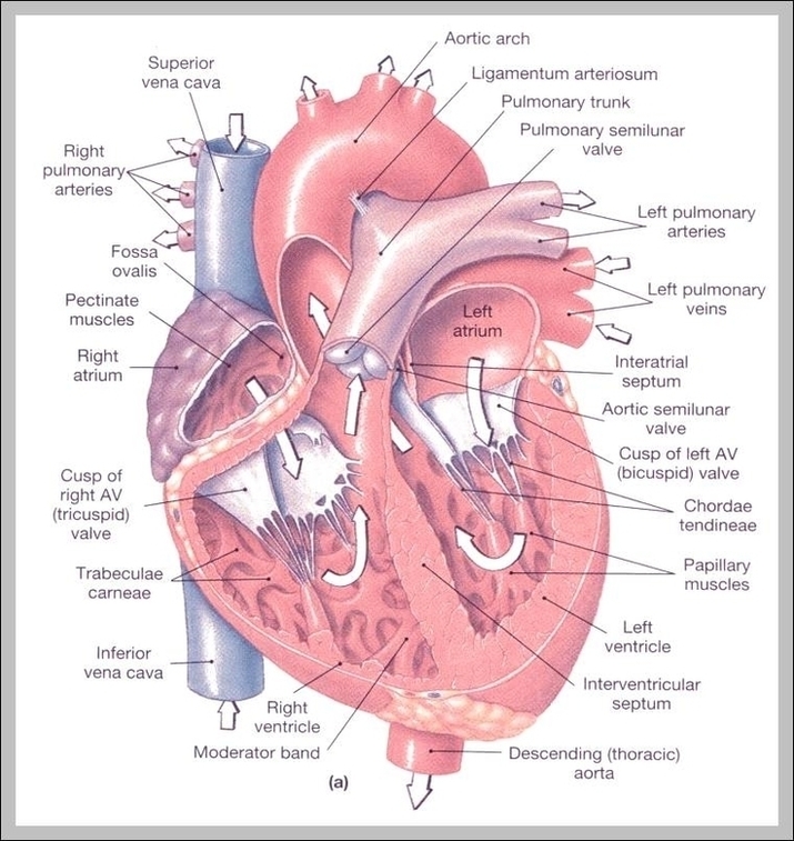
Anatomy The left auricle is a thin pouch of the heart wall located on the anterior surface of the left atrium. Its walls are only about one sixteenth of an…
Most people are familiar with the basic function of the spinal cord—how it acts as a relay that carries signals from the brain to the rest of the nervous system.…
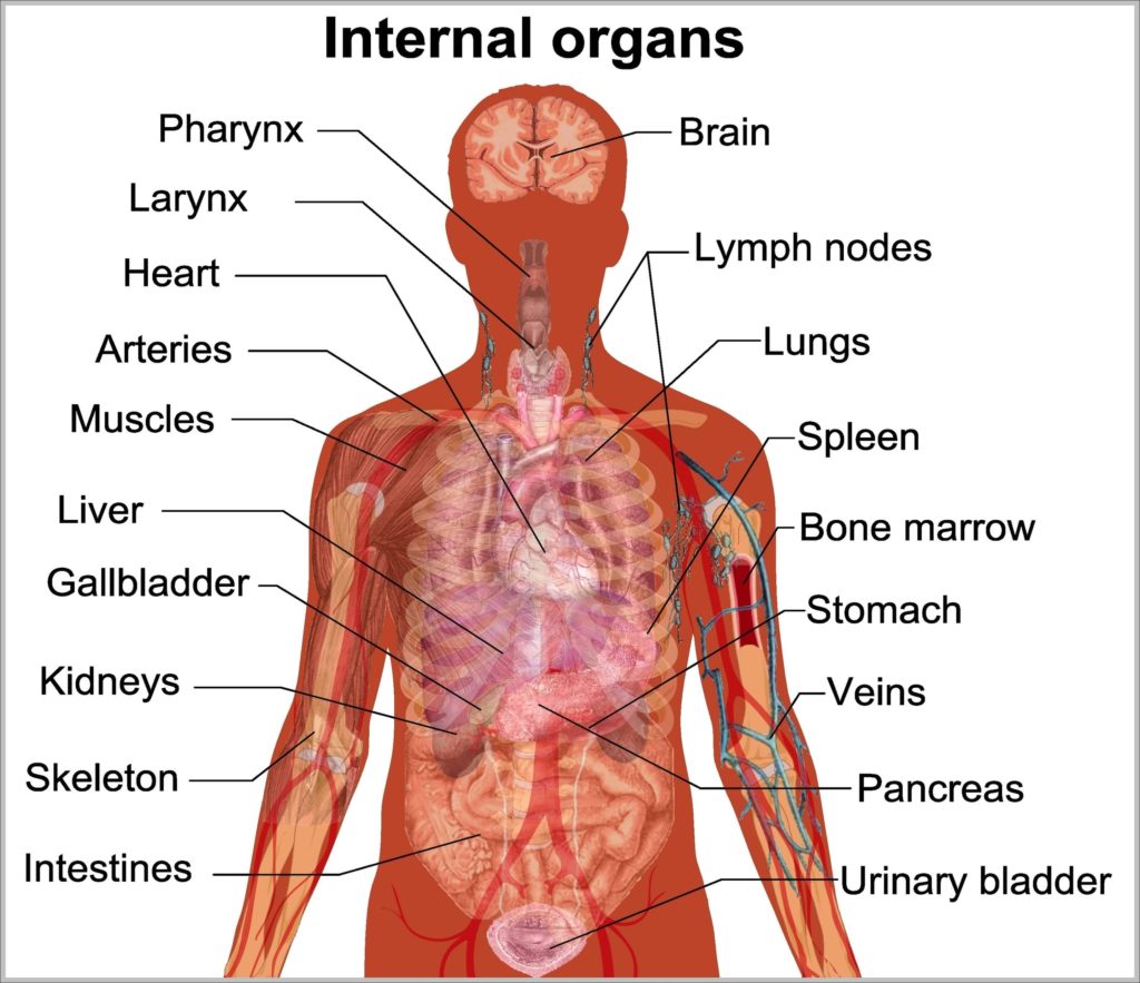
80,662 human internal organ stock photos and images available, or search for human internal organ icons or human internal organ illustrations to find more great stock photos and pictures. 7,751…
Pulmonary trunk. The pulmonary trunk or main pulmonary artery is the solitary arterial output from the right ventricle, transporting deoxygenated blood to the lungs for oxygenation. 1 Structure. The pulmonary…
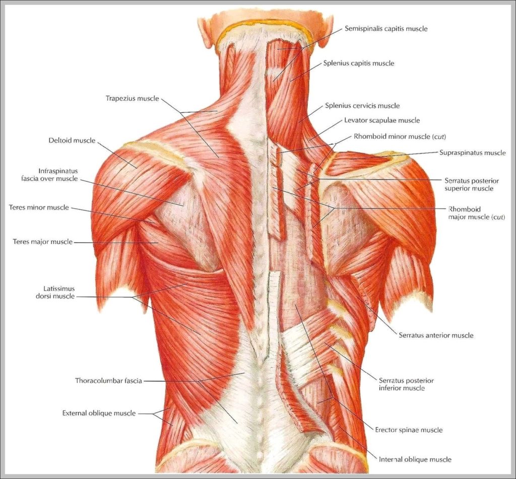
25,649 back muscle anatomy stock photos, vectors, and illustrations are available royalty-free. Best viewed on 1280 x 768 px resolution in any modern browser. This article is about Pictures Of…
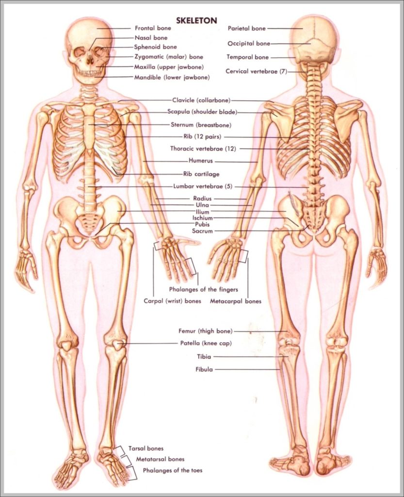
132,852 human skeleton stock photos and images available, or search for human skeleton anatomy or human skeleton vector to find more great stock photos and pictures. Male Human skeleton, four…
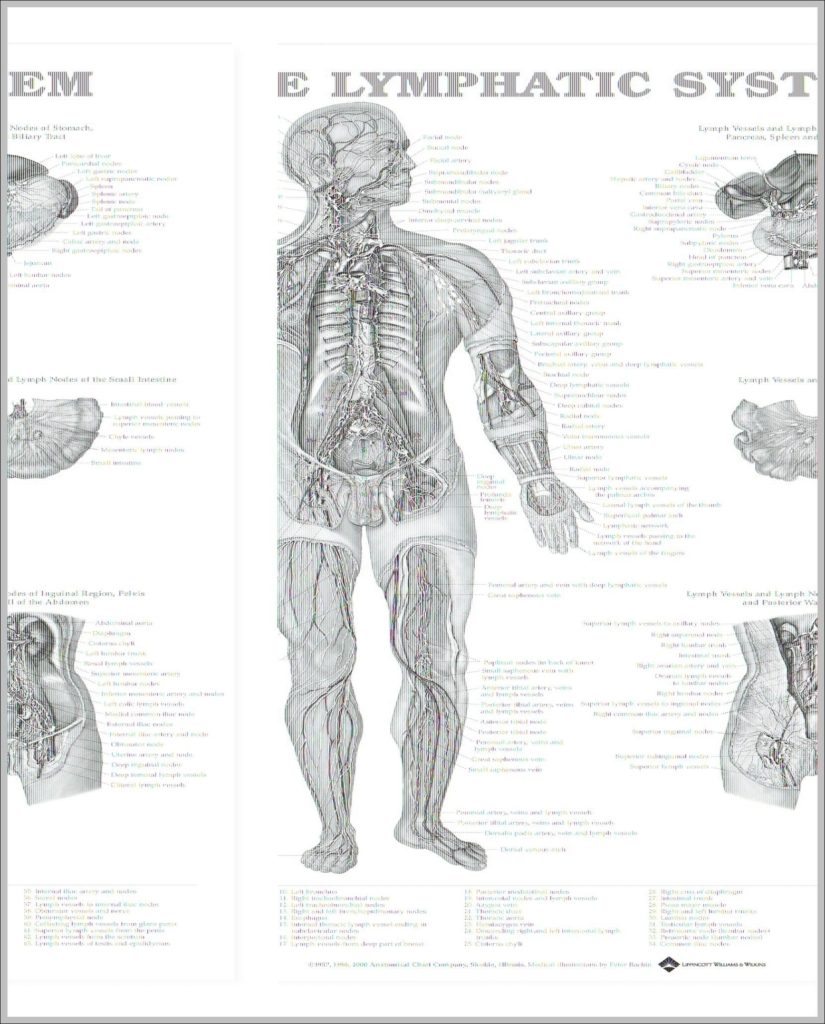
The lymphatic system consists of a conducting network of lymphatic vessels, lymphoid organs, lymphoid tissues, and the circulating lymph. 4,942 lymphatic system stock photos, vectors, and illustrations are available royalty-free.…
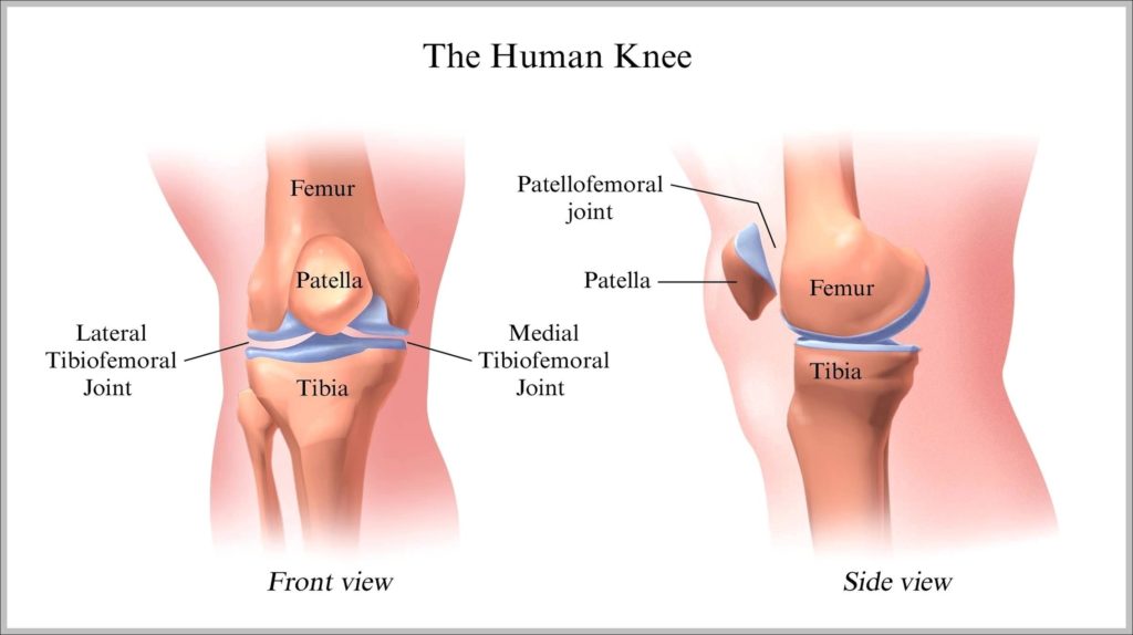
12,181 human knee stock photos and images available, or search for human knee anatomy or human knee bone to find more great stock photos and pictures. WebMD's Knee Anatomy Page…
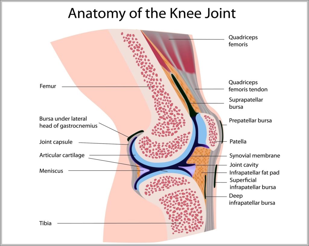
3,344 knee anatomy stock photos and images available, or search for knee anatomy illustration or human knee anatomy to find more great stock photos and pictures. Anatomy of the Knee…
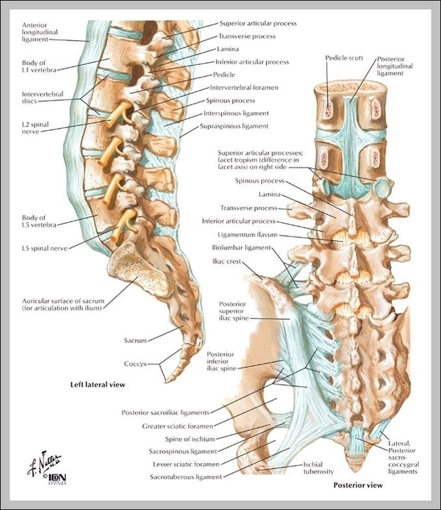
1,343 lumbar spine anatomy stock photos and images available, or search for spinal cord to find more great stock photos and pictures. Anatomical diagrams illustrate the human nervous system, especially…
27,556 human neck anatomy stock photos, vectors, and illustrations are available royalty-free. Human neck muscle anatomy. For the education Articulated human skull bone and cervical vertebrae for head and neck…
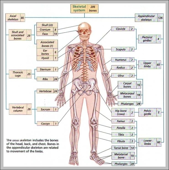
643 skeletal system stock photos are available royalty-free. Old vintage anatomy charts of the human body. Showing the skeletal system and various muscles, four figures in a row in different…