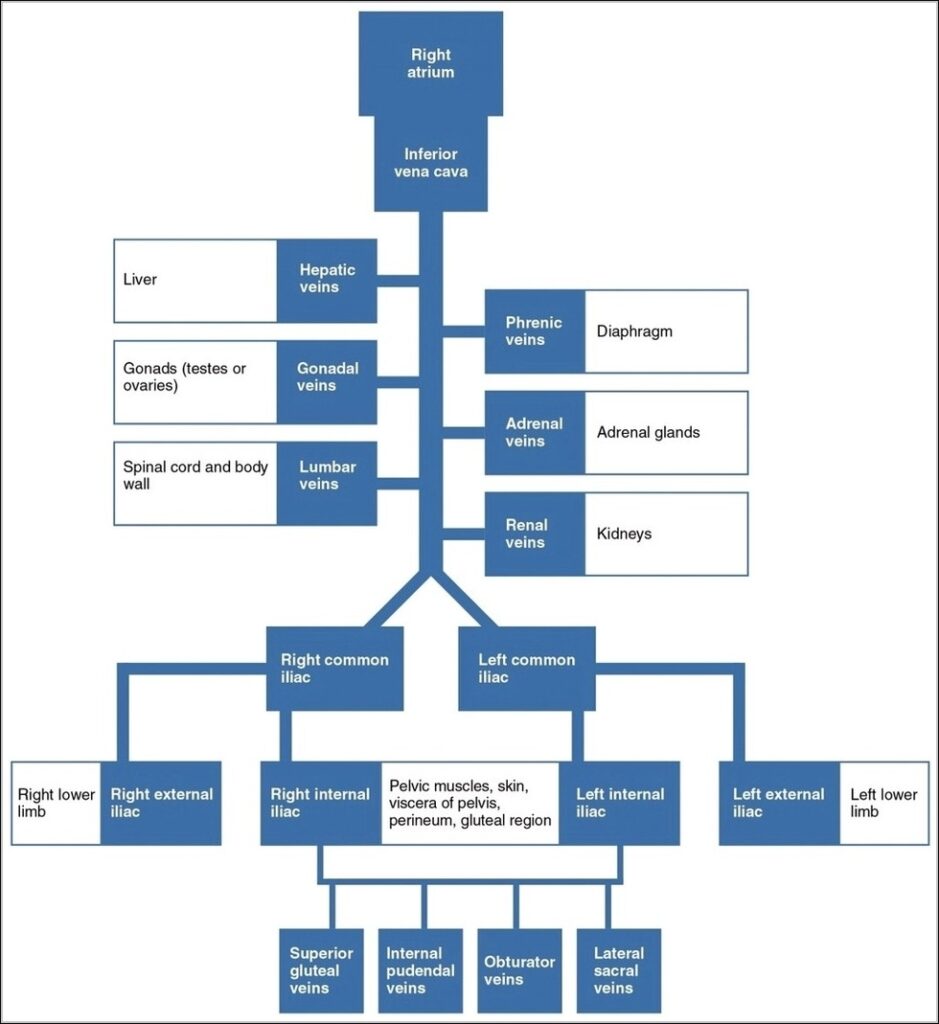
Venous blood from the body converges into two great veins: the superior and inferior vena cava. Head and neck veins drain into the brachiocephalic veins, which merge into the superior…
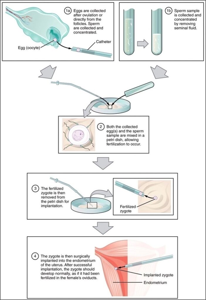
In vitro fertilization bypasses natural barriers by retrieving mature eggs from stimulated ovaries, fertilizing with sperm in labsometimes via ICSI injectionculturing embryos to blastocyst, then transferring to prepared uterus, with…
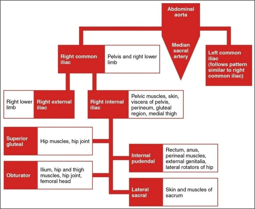
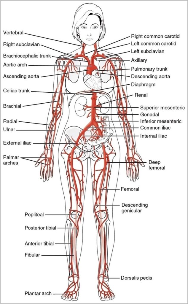
The common iliac arteries branch from abdominal aorta bifurcation at L4, each dividing into external iliac supplying lower limb via femoral and internal iliac feeding pelvis. Internal branches include superior…
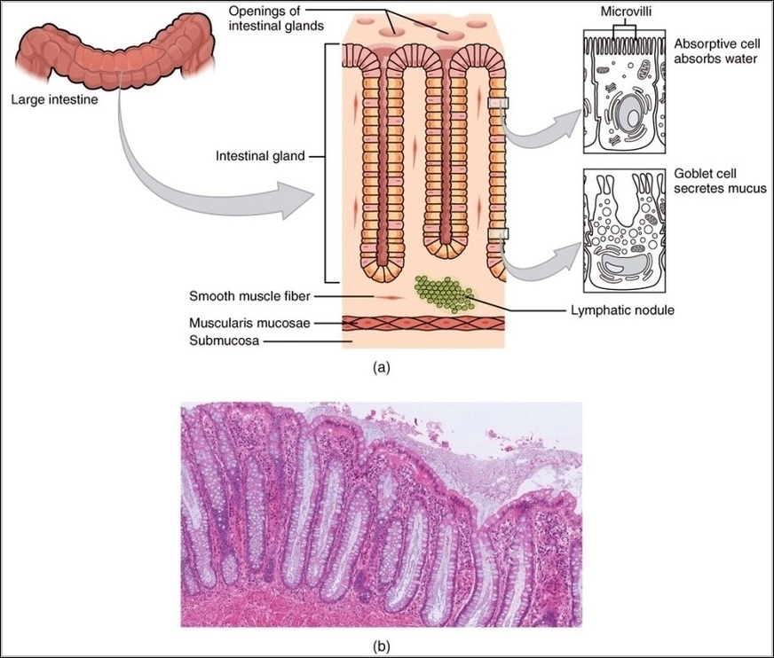
The large intestine's histology is geared toward water absorption, electrolyte balance, and waste formation, with a mucosa lacking villi but featuring deep crypts of Lieberkühn lined by columnar absorptive cells…
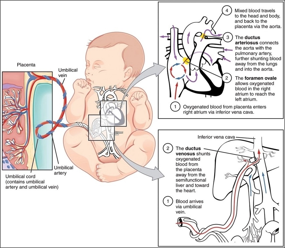
The fetal circulatory system is beautifully adapted to bypass the lungs and liver while maximizing placental exchange. Blood leaves the fetus through two umbilical arteries branching from the iliac arteries,…
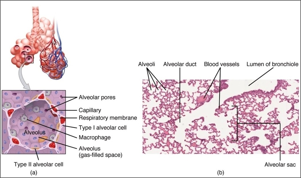
Structures of respiratory zone begin distal terminal bronchioles with occasional alveoli respiratory bronchioles more leading alveolar ducts fully walled sacs clusters sharing openings millions thin-walled pouches type I cells diffusion…
Major systemic arteries begin with ascending aorta giving coronaries, arch branching brachiocephalic, left common carotid, left subclavian, then descending thoracic supplying intercostals, abdominal continuing as celiac, mesenteric, renal, gonadal, iliacs,…
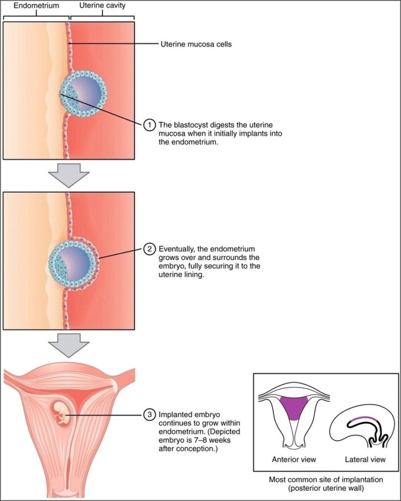
Implantation occurs about a week after fertilization when the blastocyst, having hatched from zona pellucida, adheres to endometrial lining primed by progesterone, with trophoblast cells invading to establish placental connections.…
The arterial supply to lower limbs forms a continuous chain with branches providing redundancy, starting with common femoral dividing into superficial femoral through adductor canal and profunda femoris laterally, then…
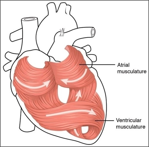
The heart's muscular wall, the myocardium, consists of specialized cardiac muscle cells arranged in spiral and circular bundles unique to each chamber. Atrial muscle is thinner and arranged in loops,…
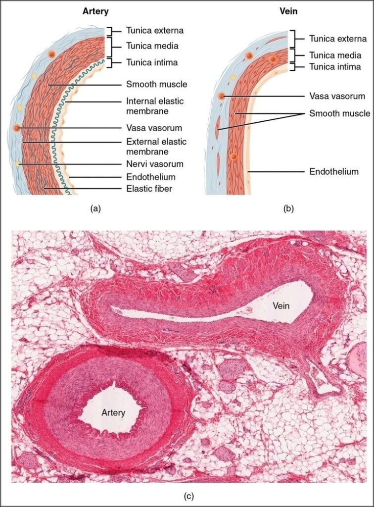
A side-by-side vascular comparison highlights the structural differences between arteries and veins. Thick muscular walls, narrow lumens, and elastic layers are shown in arteries, while veins appear thinner with wider…
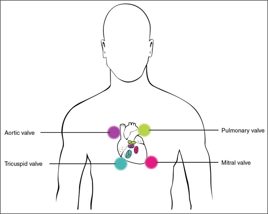
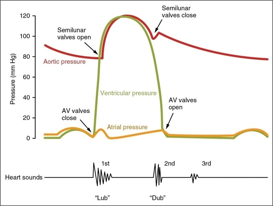
Stethoscope placement for heart sounds uses four standard sites: aortic second right intercostal sternal border high-pitched semilunar closures, pulmonic second left for same, tricuspid lower left sternal for right AV,…
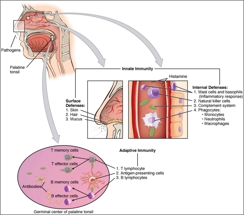
An immune system interaction diagram illustrates how innate and adaptive responses work together. Early defenses such as macrophages and inflammation are shown activating and guiding lymphocytes. This cooperation explains faster…
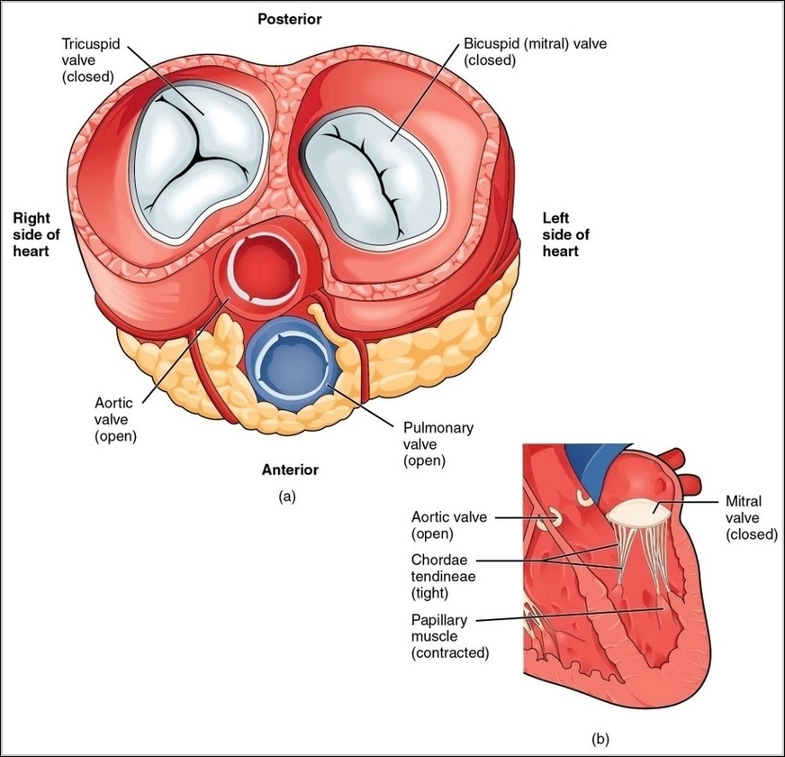
A heart cycle image showing contracted ventricles focuses on blood being forced into the aorta and pulmonary artery. Valve positions and flow direction clarify how each heartbeat moves blood efficiently…
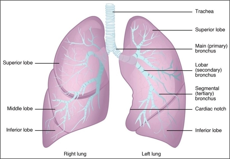
The lungs are paired spongy organs in the thoracic cavity, each divided into lobesthree on the right, two on the leftto accommodate the heart's position. The right lung is shorter…
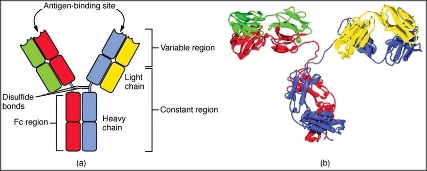
IgG antibodies consist of four polypeptide chains: two identical heavy chains and two identical light chains linked by disulfide bonds into a Y-shaped molecule. The heavy chains determine the classgamma…
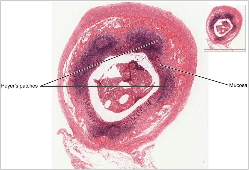
Mucosa-associated lymphoid tissue, including nodules like Peyers patches in ileum, tonsils, and appendix, forms diffuse surveillance networks beneath epithelial surfaces rich in antigens, with M cells sampling lumen contents for…
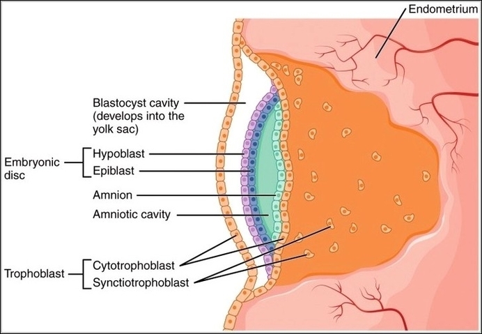
The embryonic disc with amniotic cavity and yolk sac represents the early post-implantation stage, where the bilaminar disc of epiblast and hypoblast forms, then gastrulation creates a trilaminar disc with…
A comparison of the cardiac cycle and heart sounds visually links mechanical events in the heart to what is heard through a stethoscope. The diagram aligns phases such as atrial…