Posted inDiagrams
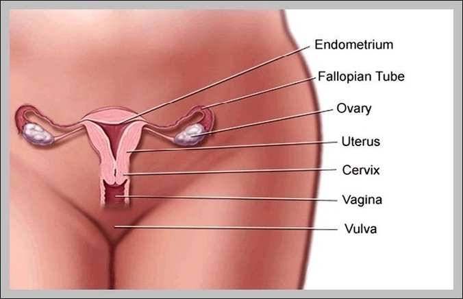
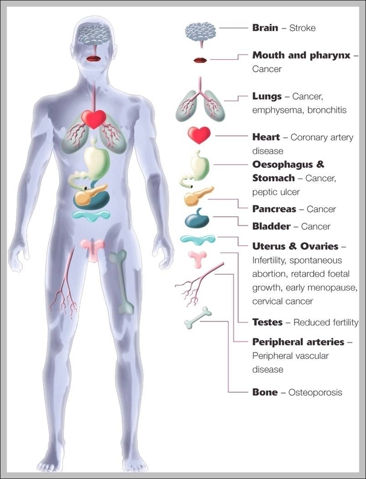
Diagram Of Human Organs In The Body Female Image
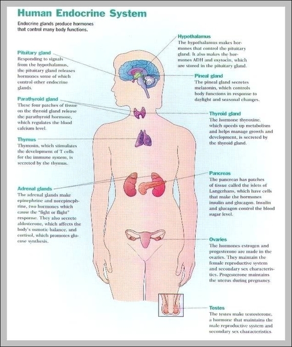
507 female body diagram stock photos and images available, or search for body silhouette or human body diagram to find more great stock photos and pictures. The Principal Glands Of…