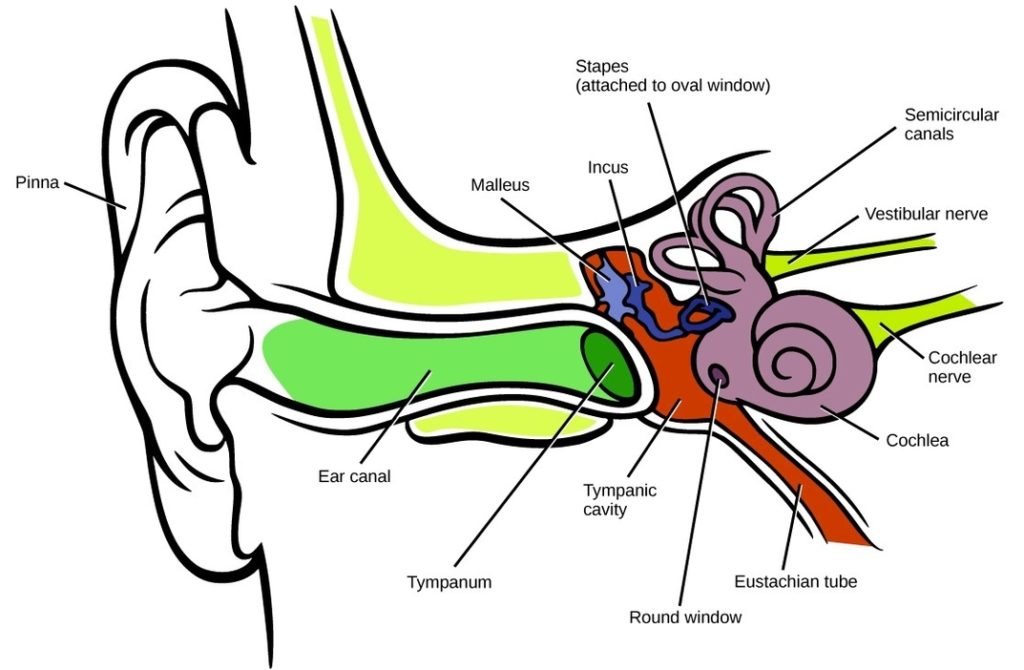
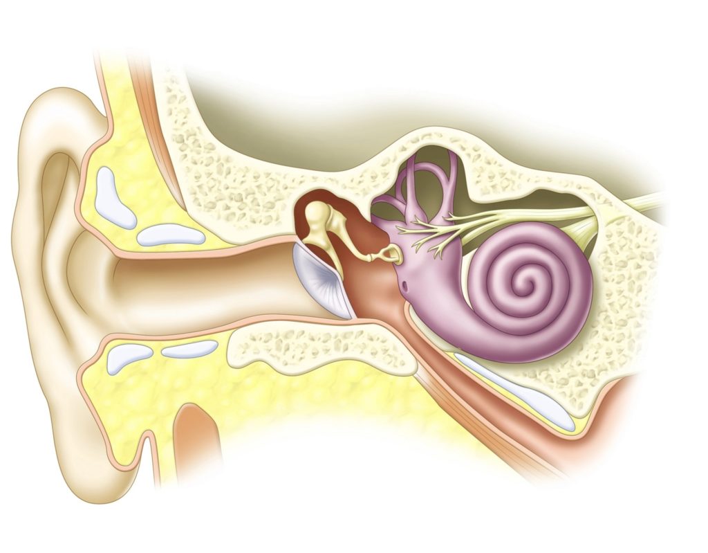
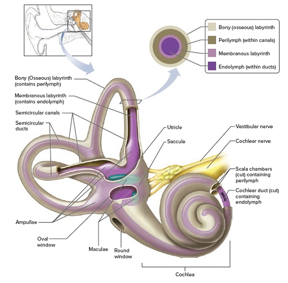
The inner ear, also known as the labyrinth, is the deepest part of your ear and plays a crucial role in hearing and maintaining balance. It consists of tiny bony…
The human ear is a complex organ that not only enables us to hear, but it also plays a key role in maintaining our body's balance. Structurally, the ear is…
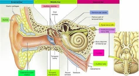
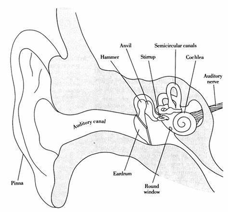
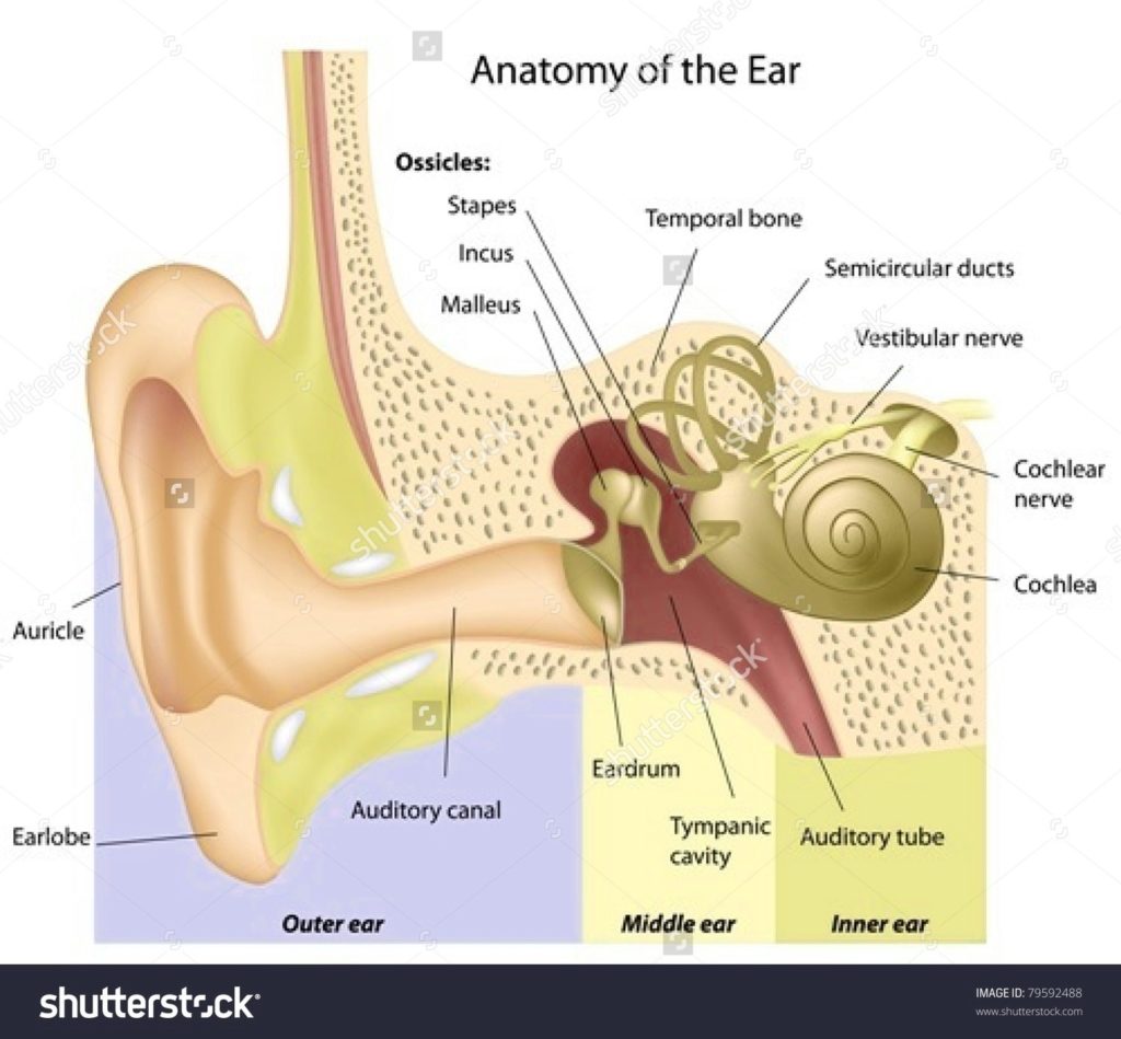
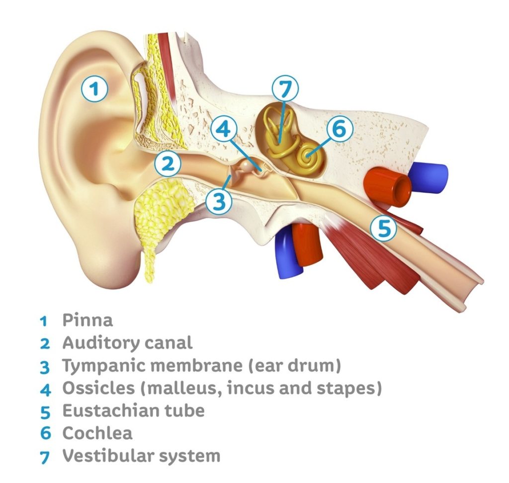
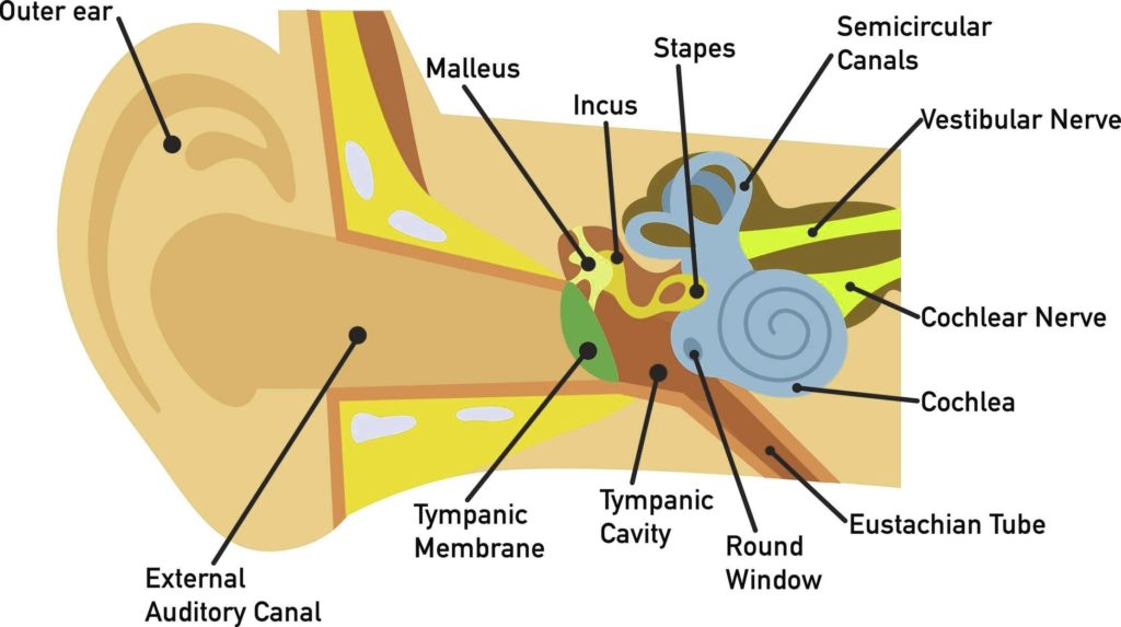
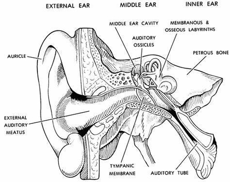
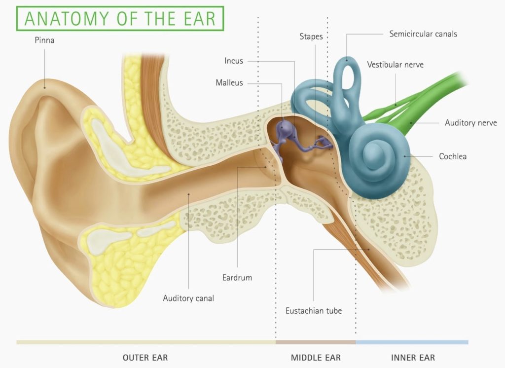
The human ear is a complex organ that serves two primary functions: hearing and maintaining balance. It is typically divided into three parts: the outer ear, the middle ear, and…
The Anatomy of the Human Ear The human ear is a complex organ that serves two primary functions: hearing and maintaining balance. It is composed of three main parts: the…
The inner ear, also known as the labyrinth, is the deepest part of the ear and plays a crucial role in hearing and maintaining balance. It consists of tiny bony…
The Anatomy of the Human Ear The human ear is a complex organ that serves two primary functions: hearing and maintaining balance. It consists of three main parts: the outer…
Inner Ear Anatomy The inner ear, also known as the labyrinth, is a complex structure responsible for hearing and balance. It is the innermost part of the ear and consists…
Human Ear Anatomy The human ear is a complex sensory organ responsible for hearing and maintaining balance. It is anatomically divided into three parts: the outer ear, middle ear, and…
The Inner Ear and Its Connection to the Brain The inner ear, also known as the labyrinth, is a complex structure that plays a crucial role in our ability to…
Ear, Nose, and Throat Anatomy Poster An Ear, Nose, and Throat (ENT) Anatomy Poster is a visual tool designed to provide a comprehensive overview of the interconnected structures involved in…
The human ear is a complex organ that serves two main functions: hearing and maintaining balance. It is typically divided into three main parts: the outer ear, the middle ear,…
The human ear is a complex organ that serves two primary functions: hearing and maintaining balance. It is anatomically divided into three distinct parts: the outer ear, the middle ear,…
Inner Ear Anatomy The inner ear, the innermost part of the ear, plays a crucial role in hearing and balance. It consists of tiny bony structures filled with fluid. As…
Human Ear Anatomy and Physiology The human ear is a complex organ that serves two primary functions: hearing and maintaining balance. It consists of three main parts: the outer ear,…
Ear Anatomy The human ear is a complex organ that serves two primary functions: hearing and maintaining balance. It is anatomically divided into three main parts: the outer ear, the…
The human ear is a complex organ that serves two main functions: hearing and maintaining balance. Here are some key parts of the ear and their functions according to Quizlet:…
The Anatomy of the Human Ear The human ear is a complex organ that serves two primary functions: hearing and maintaining balance. It is typically divided into three main parts:…
The inner ear, the innermost part of the ear, plays a crucial role in hearing and balance. It consists of tiny bony structures filled with fluid. As sound waves travel…