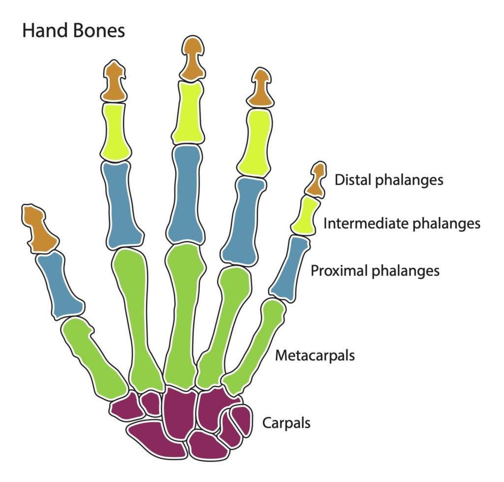
The human hand is a marvel of evolution and engineering, capable of performing a wide range of complex actions. It consists of 27 bones, divided into three categories: the carpal…
The human hand is a marvel of evolution and one of the most complex and versatile structures in the body. It consists of 27 bones that work together to provide…
Anatomy of the Left Hand The human hand, a remarkable feat of engineering and evolution, is strong enough to allow climbers to tackle any mountain, yet also sufficiently precise for…
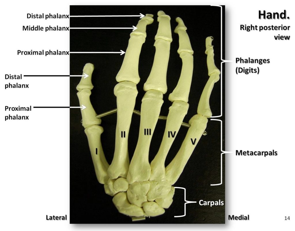
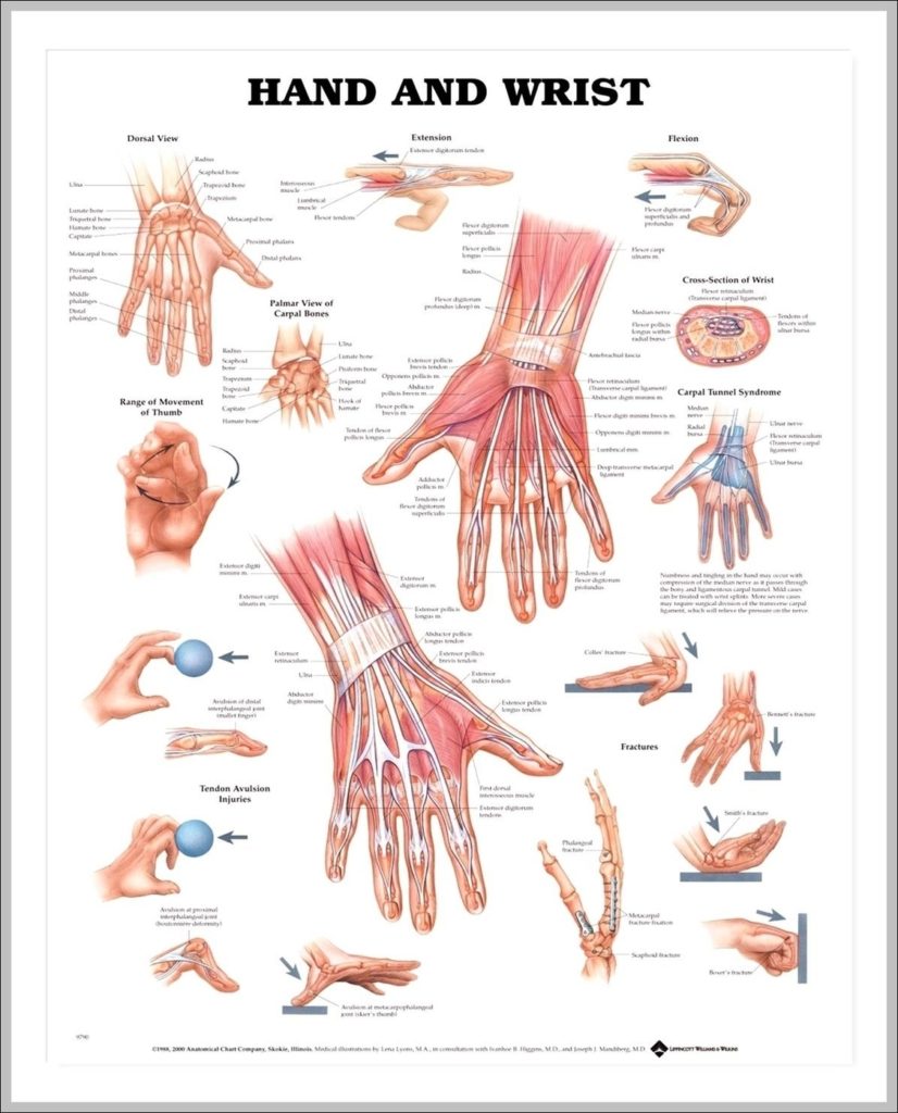
Anatomy of the Hand and Wrist: Bones, Muscles, Tendons, Nerves. The wrist links the hand to the arm. The wrist is a complex mechanical system of 8 small bones known…
The bones of the hand and wrist provide the body with support and flexibility to manipulate objects in many different ways. Each hand contains 27 distinct bones that give the…
1,317 hand bone stock photos and images available, or search for skeleton hand or human bone to find more great stock photos and pictures. Carpal, metacarpal and phalanges of the…
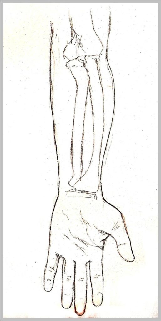
Pronation and supination of the hand: Anatomy and biomechanics Proper functioning of the hand relies on its capacity to rotate and point the palm upward (i.e. supination) or downward (i.e.…
148,045 hand anatomy stock photos, vectors, and illustrations are available royalty-free. Picture of Hand. There are 28 phalanges (finger bones) and 10 metacarpal bones. Each finger has 3 phalanges and…
1,317 hand bone stock photos and images available, or search for skeleton hand or human bone to find more great stock photos and pictures. Carpal, metacarpal and phalanges of the…
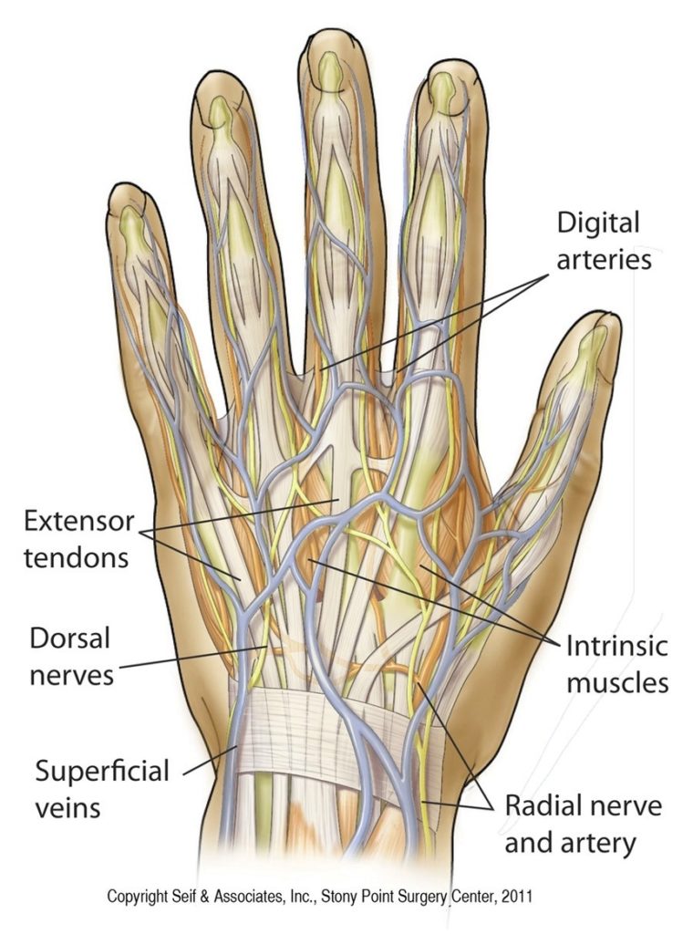
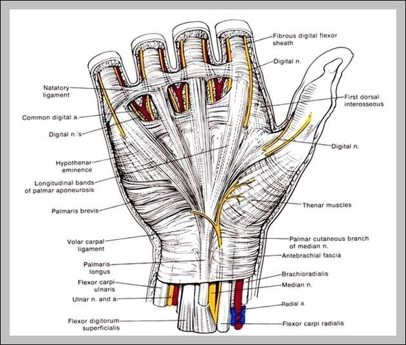
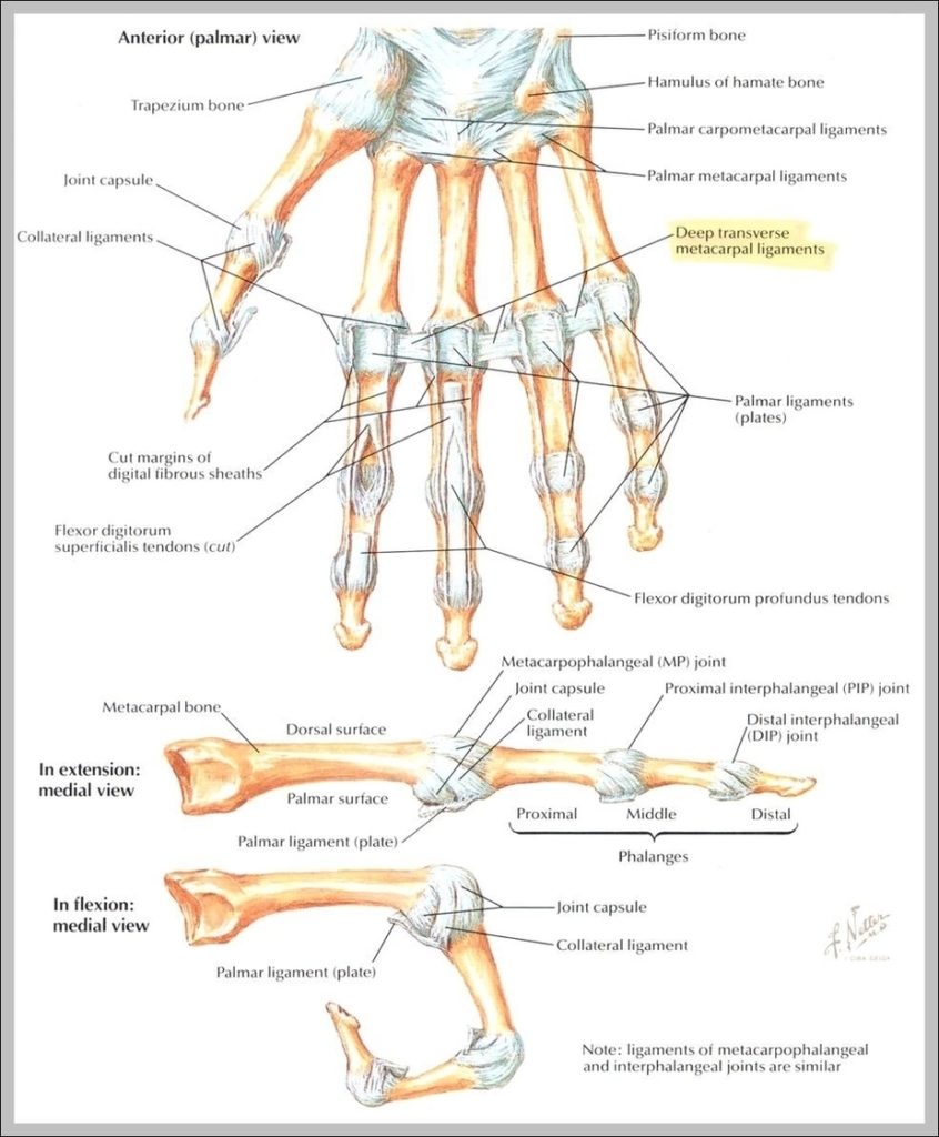
Ligaments. There are many ligament of the hand that are made up of tough bands of fibrous tissue. As there are many small bones and joints in the hand, there…
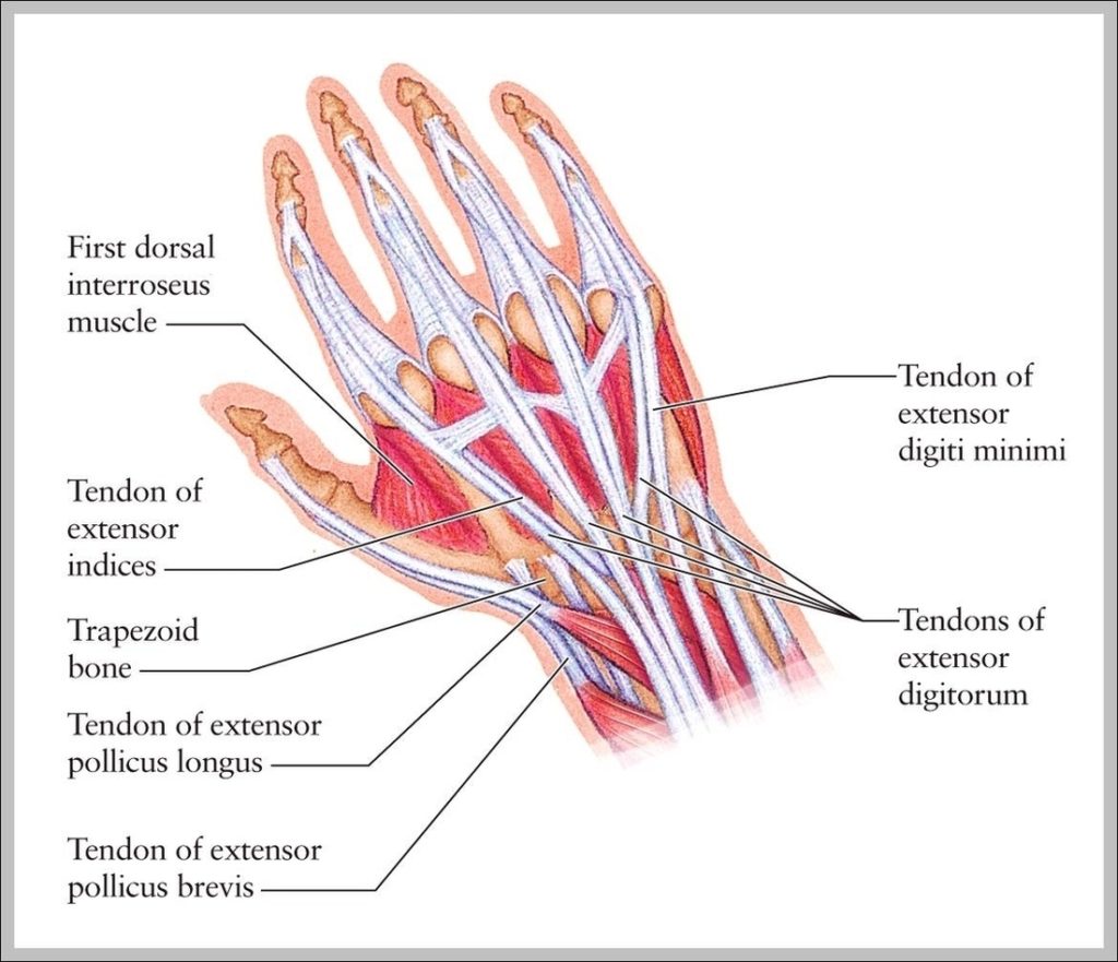
324 hand anatomy tendons stock photos and images available, or start a new search to explore more stock photos and images. Vintage anatomical color illustration of the musculature of the…
In purely anatomical terms, only those ligaments that connect the bones of the hand to each other are properly called hand ligaments. The ligaments of the fingers and those that…
148,045 hand anatomy stock photos, vectors, and illustrations are available royalty-free. To understand the anatomy of the hand we first must understand the anatomy of the forearm and wrist. The…
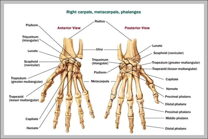
The wrist links the hand to the arm. The wrist is a complex mechanical system of 8 small bones known as the carpal bones. The carpal bones are arranged in…
The wrist tendons are: Flexor carpi radialis: This tendon is one of two tendons that bend the wrist. It attaches to the base of the second and third hand bones.…
In purely anatomical terms, only those ligaments that connect the bones of the hand to each other are properly called hand ligaments. The ligaments of the fingers and those that…