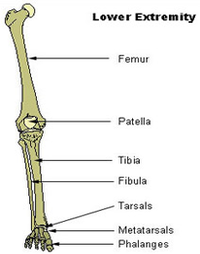
Key facts about the lower extremity Hip and pelvis Bones: hip bones, saccrum, coccyx Hip jo … Thigh Bones: femur Joints: hip and knee Muscle … Knee Bones: tibia, fibula, patella Type: hing … Leg Bones: tibia, fibula Joints: knee and an … Ankle and foot Ankle joint: hinged joint capable of …
Each lower extremity artery is visible with an accompanying vein, extending from the iliac artery to the popliteal artery. The anterior tibial artery, the posterior tibial artery, and the peroneal artery are seen with two homonymous veins. The overall anatomy of the arteries in the lower extremities is shown on CT angiography in Fig. 1.
The lower leg is a major anatomical part of the skeletal system. Together with the upper leg, it forms the lower extremity. It lies between the knee and the ankle, while the upper leg lies between the hip and the knee. The lower leg contains two major long bones, the tibia and the fibula, which are both very strong skeletal structures.

lower extremity diagram
Posted inDiagrams
