The human skull, a marvel of biological engineering, serves as the framework for the face and a protective case for the brain. It consists of 22 bones, divided into two main parts: the cranium (or neurocranium) and the facial skeleton (or viscerocranium)?.
Cranium
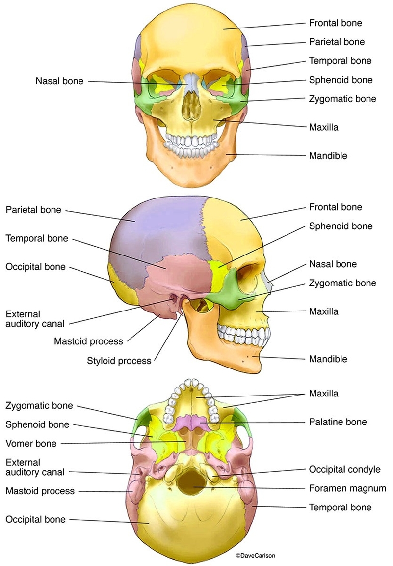
The cranium, which encloses and protects the brain, is composed of eight bones?:
1. Frontal Bone: Forms the forehead and the roofs of the orbits (eye sockets).
2. Parietal Bones (2): Form the sides and roof of the cranium.
3. Temporal Bones (2): House the structures of the ears and contribute to the sides and base of the skull.
4. Occipital Bone: Forms the back and base of the cranium, containing the foramen magnum through which the spinal cord passes.
5. Sphenoid Bone: Forms part of the base of the skull and parts of the floor and sides of the orbits.
6. Ethmoid Bone: Contributes to the walls of the orbits, the roof and walls of the nasal cavity, and the nasal septum.
Facial Skeleton
The facial skeleton comprises 14 bones?:
1. Maxillae (2): Form the upper jaw, the anterior roof of the mouth, the floors of the orbits, and the sides and floor of the nasal cavity.
2. Palatine Bones (2): Form the posterior part of the hard palate and the floor of the nasal cavity.
3. Zygomatic Bones (2): Form the cheeks and the lateral walls and floors of the orbits.
4. Lacrimal Bones (2): Contribute to the walls of the orbits and house the lacrimal sacs, which help drain tears into the nasal cavity.
5. Nasal Bones (2): Form the bridge of the nose.
6. Inferior Nasal Conchae (2): Form part of the interior of the nose.
7. Vomer: Forms the inferior part of the nasal septum.
8. Mandible: Forms the lower jaw and is the only moveable bone of the skull.
The skull also contains several sutures, which are joints that hold the bones together. The most significant are the coronal suture (between the frontal and parietal bones), the sagittal suture (between the two parietal bones), and the lambdoidal suture (between the occipital and parietal bones)?.
Human Skull Model
A human skull model is a replica of the human skull, often used for educational purposes. These models are anatomically correct, providing detailed representations of the skull’s structure. They can be used to study the interior and exterior anatomical structures of the human skull in great detail. Some models are designed with detachable parts, such as the mandible or individual bones, to allow for a more in-depth examination. These models are invaluable tools for medical students, professionals, and educators alike, offering a hands-on approach to studying the complex anatomy of the human skull.