The human leg, a marvel of biological engineering, is a complex structure composed of numerous bones. These bones are designed to withstand daily strain, absorb force, and provide flexibility for balance and movement.
Upper Leg Bone
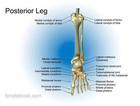
The upper leg or thigh houses the femur, the longest and strongest bone in the body. The femur forms a ball-and-socket joint with the hip bone at its proximal end and two condyles at the distal end form the knee joint with the lower leg bones.
The Knee
The knee is a hinge joint with the patella, or kneecap, covering the joint. The patella connects with the upper and lower leg bone through muscles and ligaments.
Lower Leg Bones
The region between the knee and the ankle consists of two long bones:
1. Tibia: The longer and thicker of the two bones, located medially in the lower leg. The weight-bearing bone articulates with the femur at its superior end and the tarsals at its inferior end.
2. Fibula: Located laterally, it articulates with the tibia but does not bear any of the bodys weight directly. It serves as an essential point of attachment for various leg muscles.
Foot Bones
The foot consists of several small bones that work together to distribute the bodys weight as we walk, run, dance, or do any movement with our foot:
1. Tarsals: A group of 7 bones in each foot that allow minor adjustments to the foot position when we stand or walk. These include the calcaneus (heel bone), talus, navicular bone, medial cuneiform bone, intermediate cuneiform bone, lateral cuneiform bone, and cuboid bone.
2. Metatarsals: There are 5 metatarsals in each foot, forming the forefoot.
3. Phalanges of the foot (toe bones): These tiny bones articulate with the metatarsals and then form the toes. There are 14 phalanges in each foot, providing support and allowing for various foot movements