A hinge joint is a common class of synovial joint that includes the ankle, elbow, and knee joints. Hinge joints are formed between two or more bones where the bones can only move along one axis to flex or extend.
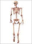
The elbows, the knees, and the middle and end joints of the fingers are hinge joints. Hinge joints are prone to injury when lateral forces are applied to the joint. Many knee injuries occur in this manner, according to the National Institute of Arthritis and Musculoskeletal and Skin Diseases.
The elbows, the knees, and the middle and end joints of the fingers are hinge joints. Hinge joints are prone to injury when lateral forces are applied to the joint. Many knee injuries occur in this manner, according to the National Institute of Arthritis and Musculoskeletal and Skin Diseases.

Hinge Joints In The Body Image
Posted inDiagrams
Hinge Joints In The Body Image
Post navigation
Previous Post
 Skeletal Pictures Image
Next Post
Image Of Human Body Image