Heart Anatomy
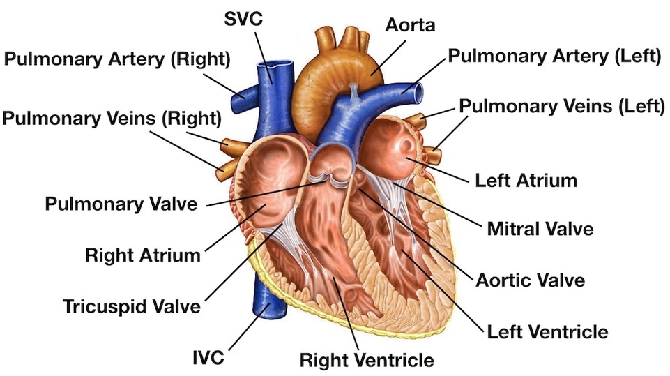
The heart is a muscular organ that pumps blood around the body by circulating it through the circulatory/vascular system. It is found in the middle mediastinum, wrapped in a two-layered serous sac called the pericardium. The heart is shaped as a quadrangular pyramid, and orientated as if the pyramid has fallen onto one of its sides so that its base faces the posterior thoracic wall, and its apex is pointed toward the anterior thoracic wall.
urfaces and Borders
The heart has five surfaces: base (posterior), diaphragmatic (inferior), sternocostal (anterior), and left and right pulmonary surfaces. It also has several margins: right, left, superior, and inferior. The right margin is the small section of the right atrium that extends between the superior and inferior vena cava. The left margin is formed by the left ventricle and left auricle. The superior margin in the anterior view is formed by both atria and their auricles. The Inferior margin is marked by the right ventricle.
Chambers
Inside, the heart is divided into four heart chambers: two atria (right and left) and two ventricles (right and left). The right atrium and ventricle receive deoxygenated blood from systemic veins and pump it to the lungs, while the left atrium and ventricle receive oxygenated blood from the lungs and pump it to the systemic vessels which distribute it throughout the body.
Valves
The heart has four valves: Tricuspid, Pulmonary, Mitral, and Aortic valves. These valves ensure that blood flows in the correct direction through the heart, preventing backflow.
Blood Supply
The heart’s blood supply is primarily provided by the right and left coronary arteries. The right coronary artery has several branches, including the sinuatrial nodal branch, right marginal branch, atrioventricular nodal branch, and posterior interventricular branch. The left coronary artery has two main branches: the circumflex branch and the anterior interventricular branch.
Great Vessels
The great vessels that originate from the heart radiate their branches to the head and neck, the thorax and abdomen, and the upper and lower limbs. These include the aorta, the superior and inferior vena cavae, and the pulmonary artery.
Conclusion
The heart’s complex structure and function make it a vital organ for life. Its chambers, valves, and blood vessels work together to pump blood throughout the body, supplying oxygen and nutrients to every cell. Understanding the anatomy of the heart is crucial for understanding how the circulatory system works and how diseases can affect this important organ..