
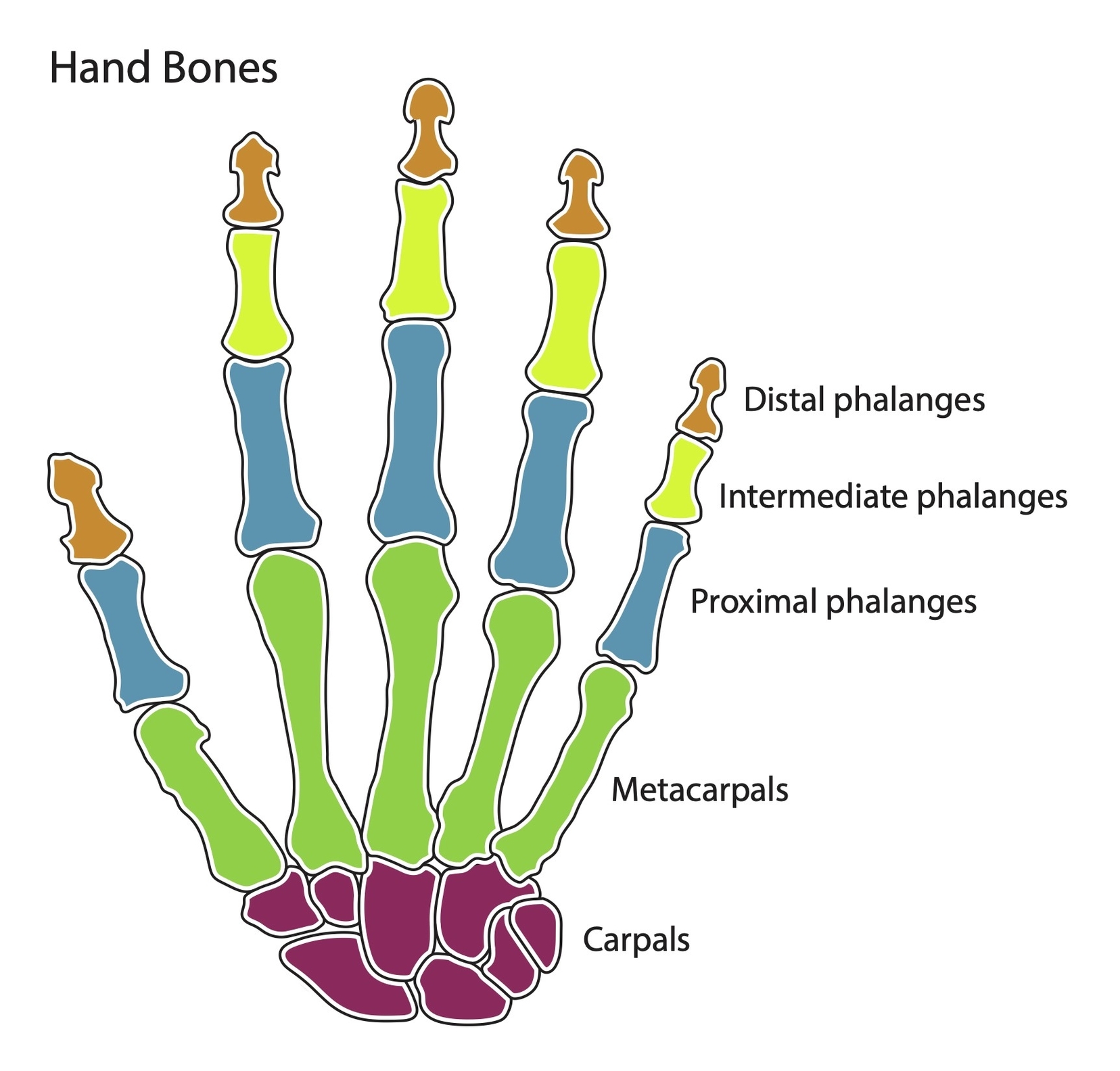
The human hand is a marvel of evolution and engineering, capable of performing a wide range of complex actions. It consists of 27 bones, divided into three categories: the carpal bones, the metacarpal bones, and the phalanges.
Carpal Bones
The carpal bones are a set of eight irregularly shaped bones located in the wrist area. They are organized into two rows: proximal and distal. The proximal row, from lateral to medial, consists of the scaphoid, lunate, triquetrum, and pisiform. The distal row, also from lateral to medial, includes the trapezium, trapezoid, capitate, and hamate. These bones form an arch in the coronal plane, creating the carpal tunnel.
Metacarpal Bones
There are five metacarpal bones, each associated with a digit. They articulate proximally with the carpals and distally with the proximal phalanges. Each metacarpal consists of a base, shaft, and a head. The medial and lateral surfaces of the metacarpals are concave, allowing attachment of the interossei muscles.
Phalanges
The phalanges are the bones of the fingers. Each finger has three phalanges: proximal, middle, and distal. The thumb, however, has only two phalanges.
Muscles and Tendons
The hand’s range of motion is not solely due to its bones. The forearm muscles, projecting tendons towards the hand via the wrist, are major contributors. The hand also has intrinsic muscles, including the thenar group (abductor pollicis brevis, flexor pollicis brevis, opponens pollicis), the hypothenar group (abductor digiti minimi, flexor digiti minimi, opponens digiti minimi), and the metacarpal group (lumbricals, palmar interossei, dorsal interossei).
Nerves and Blood Supply
The median nerve and its branches predominantly supply the thenar muscles, while the radial nerve provides cutaneous innervation along the outside of the thumb. The ulnar nerve and its branches innervate the hypothenar and metacarpal groups. The hand’s blood supply originates from the radial and ulnar arteries, which include various branches.
Clinical Relevance
Understanding the hand’s anatomy is crucial in medicine. For instance, the scaphoid bone is the most commonly fractured carpal bone, typically by falling on an outstretched hand. A fracture can interrupt the blood supply to the proximal part of the scaphoid bone, leading to avascular necrosis.
In conclusion, the hand’s intricate structure of bones, muscles, and neurovascular structures allows for its remarkable strength and precision. Its complexity underscores the importance of understanding its anatomy, whether for medical purposes or simply to appreciate the marvels of human evolution..
