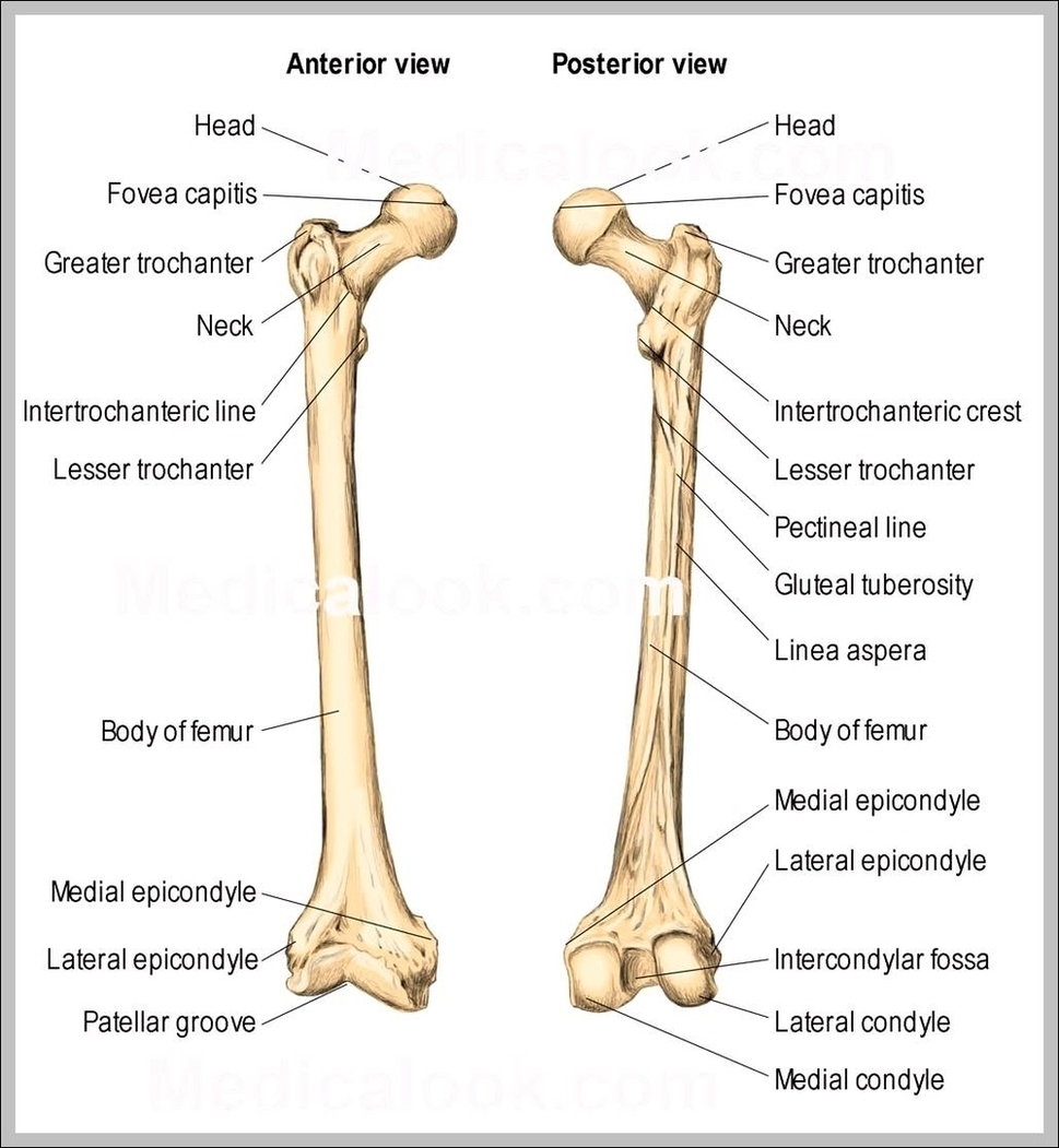
The anatomy of the femur can be divided into proximal, central, distal, and posterior parts. We will use a color-coded labeled diagram to walk through the anatomy of the femur and the different parts of the bone. By the end of this post, you will be able to label the anatomical features shown on the diagram below.
4,436 femur stock photos and images available, or search for femur bone or femur fracture to find more great stock photos and pictures. Femur, Paired bone forming the skeleton of the thigh, between the hip and knee joints.
4,436 femur stock photos and images available, or search for femur bone or femur fracture to find more great stock photos and pictures. Femur, Paired bone forming the skeleton of the thigh, between the hip and knee joints.

Femur Anatomy Diagram Image
Posted inDiagrams
