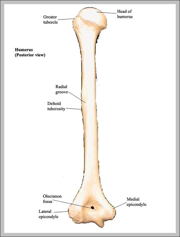
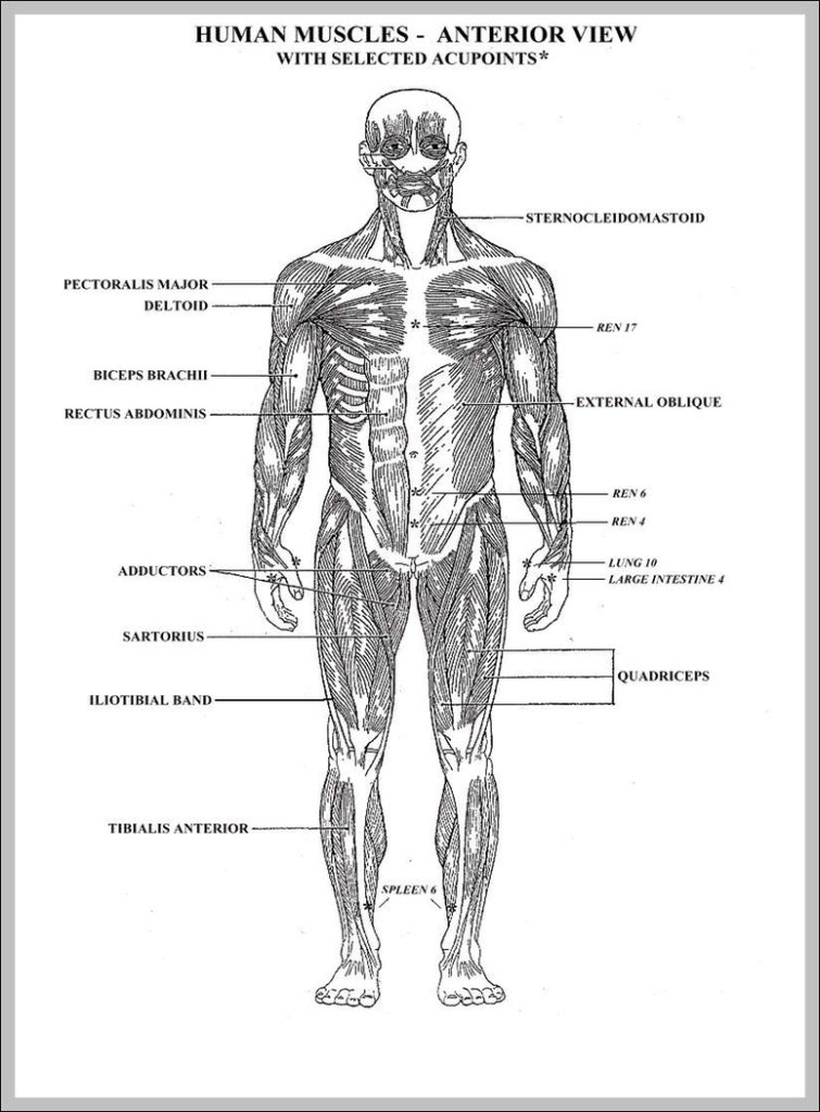
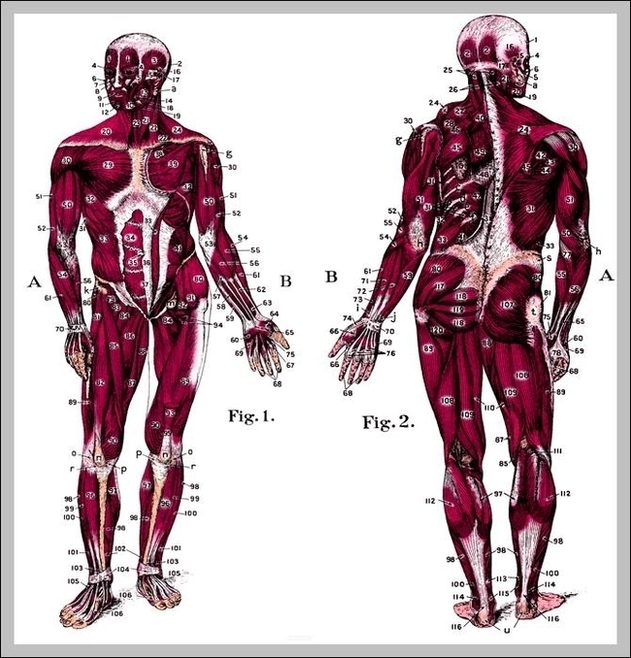
List of Upper Body Muscles 1 Neck and Shoulders. The neck primarily consists of the sternocleidomastoid and the splenius muscles, which act to flex and extend the neck, respectively. 2…
1,777 nervous system diagram stock photos and images available, or search for central nervous system diagram to find more great stock photos and pictures. Diagram of nerve reflexes: stimulus running…
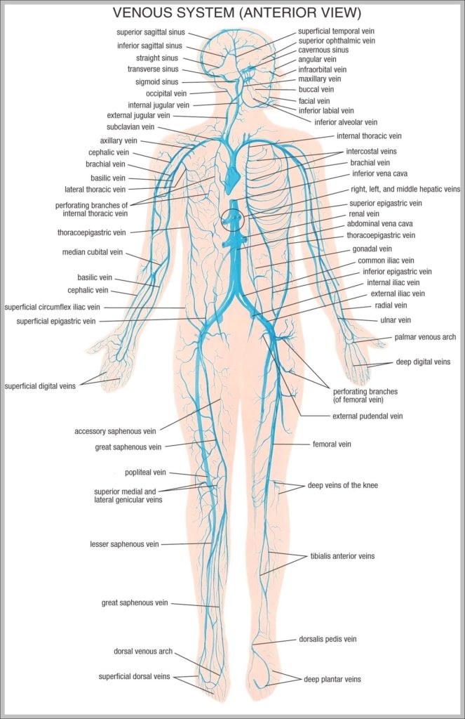
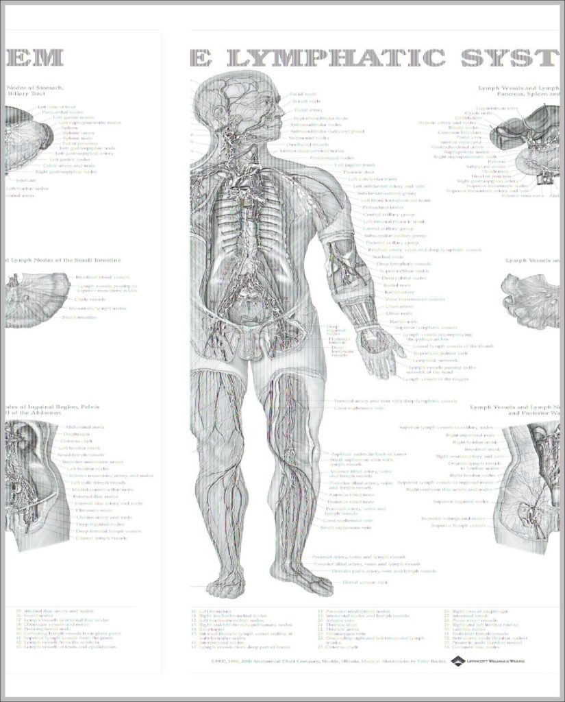
The thoracic duct carries chyle, a liquid containing both lymph and emulsified fats, rather than pure lymph. It also collects most of the lymph in the body other than from…
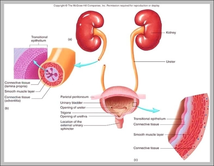
The ureter is a tube that carries urine from the kidney to the urinary bladder. There are two ureters, one attached to each kidney. ... Duplication of the ureter: a…
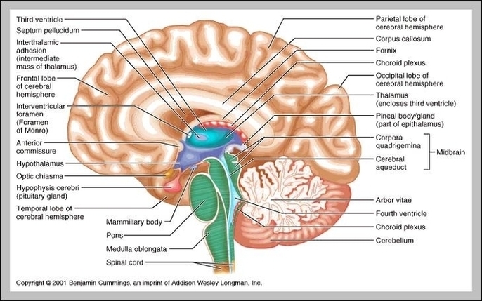
10,526 brain anatomy stock photos and images available, or search for human brain anatomy or brain anatomy illustration to find more great stock photos and pictures. WebMD's Brain Anatomy Page…
WebMD's Intestines Anatomy Page provides a detailed image and definition of the intestines. Learn about its parts, location in the body, function, and conditions that affect the intestines. home image…
The segmental conception of pulmonary anatomy is not new, but has become increasingly important in recent years. A bronchopulmonary segment is defined as that area of lung supplied by a…
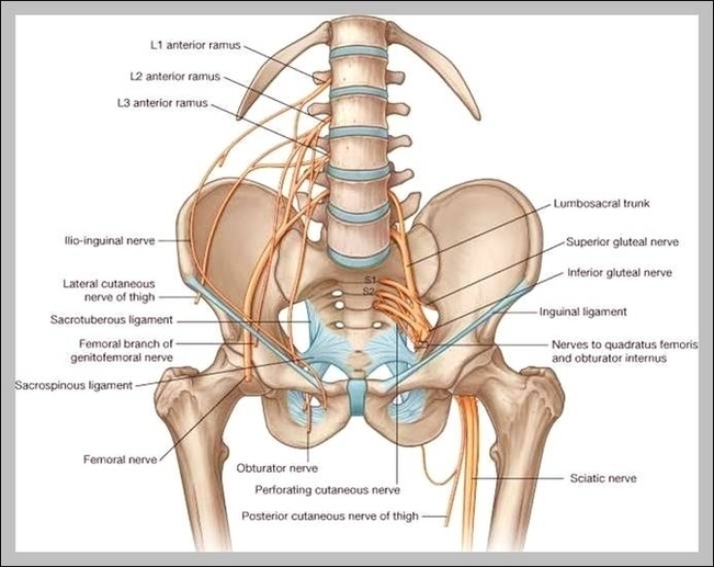
An important group of muscles in the pelvis is the pelvic floor. The pelvic floor muscles provide foundational support for the intestines and bladder. They also help the anus function.…
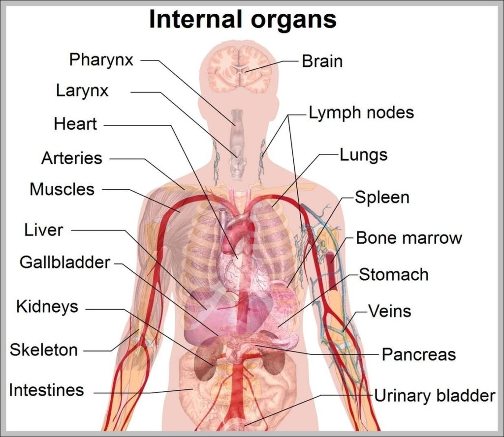
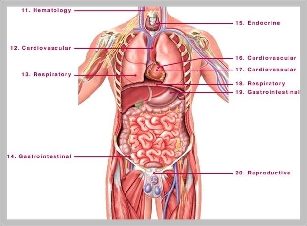
Here are a number of highest rated Location Of Major Organs Human Body pictures on internet. We identified it from well-behaved source. Its submitted by processing in the best field.…
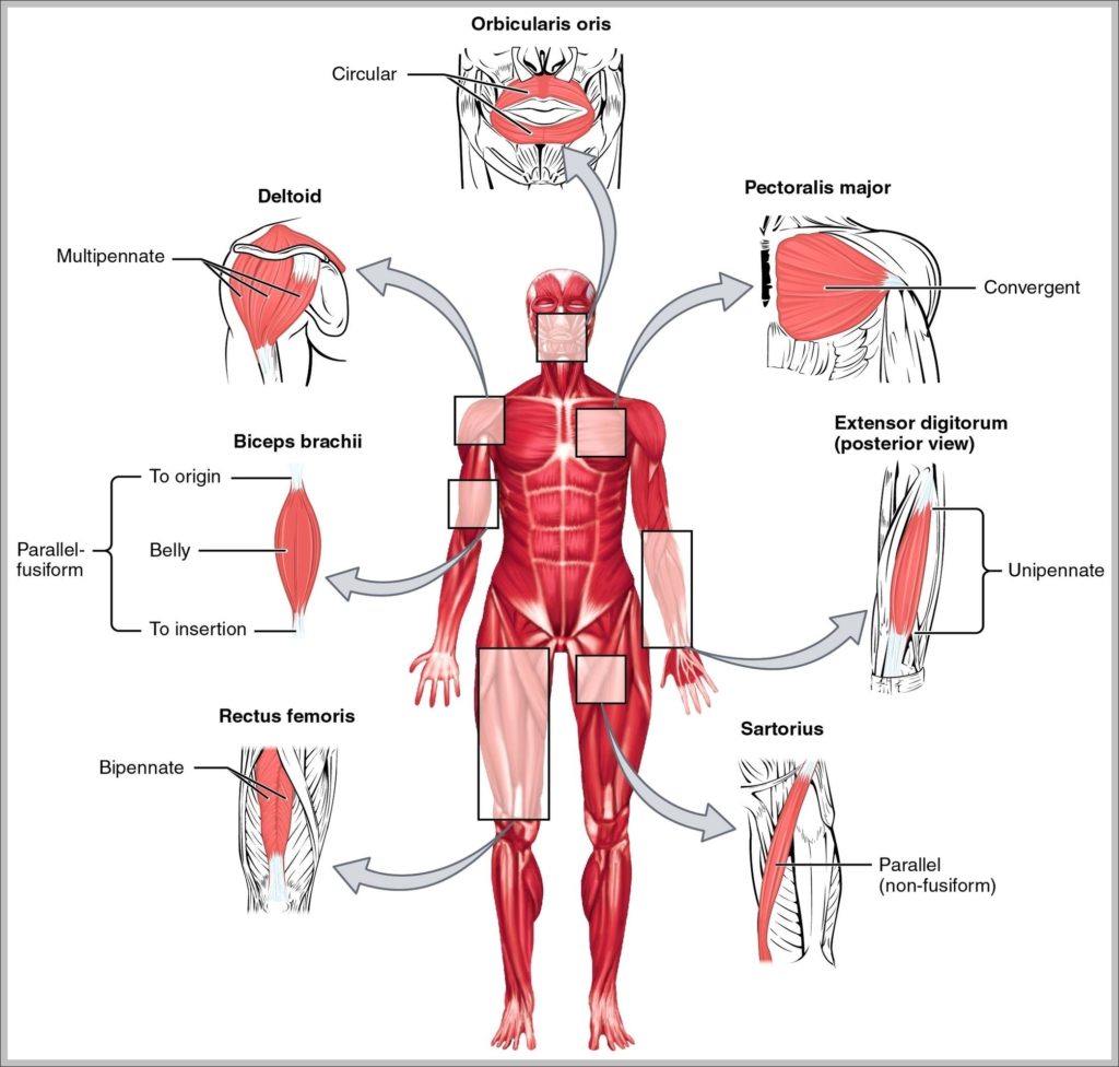
98 muscular system labeled diagram stock photos and images available, or start a new search to explore more stock photos and images. Frontal view of the muscular system of the…
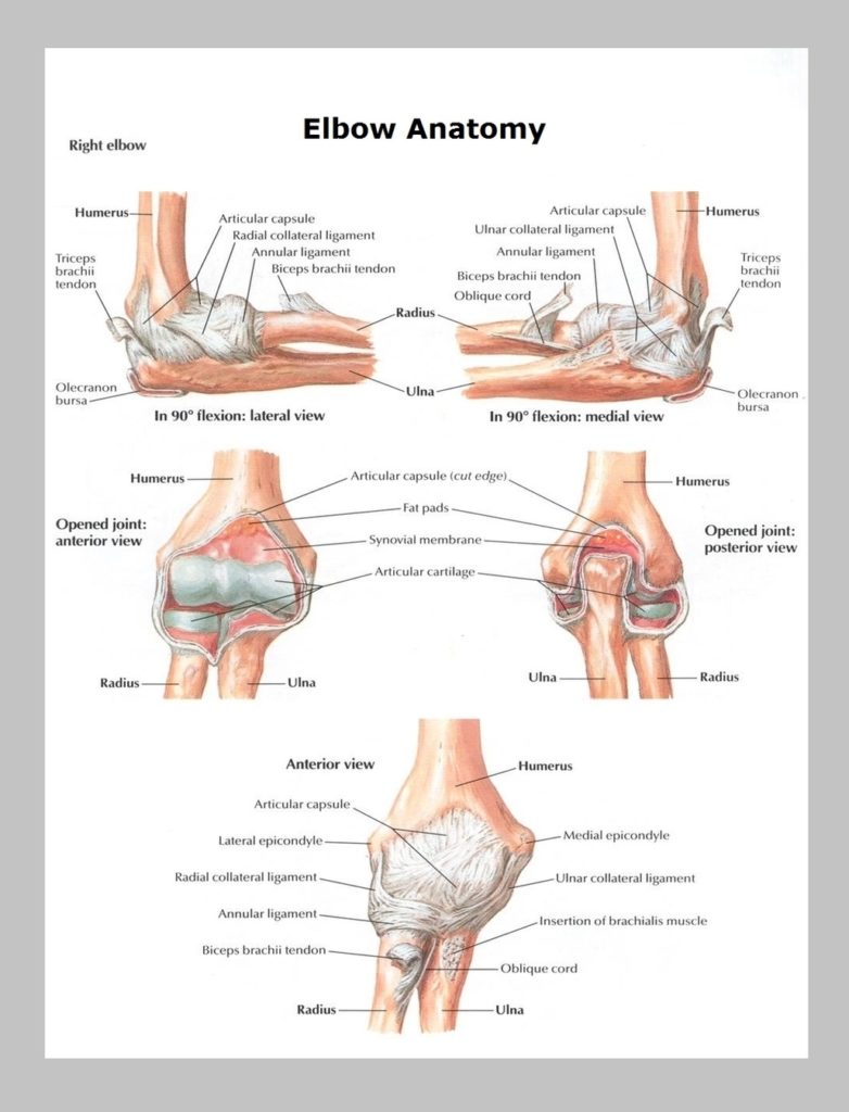
The elbow is one of the largest joints in the body. In conjunction with the shoulder joint and wrist, the elbow gives the arm much of its versatility, as well…
2,169 lymphatic system stock photos and images available or search for lymphatic system diagram or lymphatic system illustration to find more great stock photos and pictures. Illustration of a thyroid…
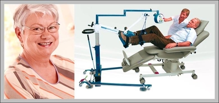
Training programs prepare nurses to additionally pursue the title of certified hemodialysis nurse. Depending on the type of dialysis machine you will use, the training program lasts for about 4…
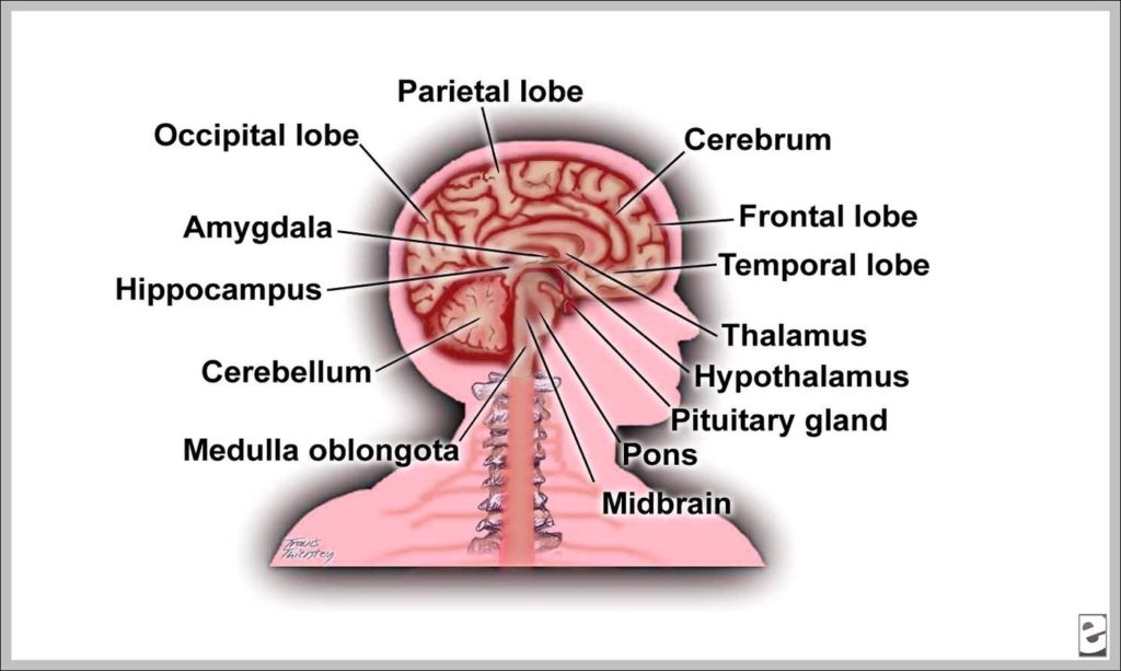
Before we have a look at the brain diagram, it is important to go through a few facts about the brain and its function. This will help you understand the…
The Wikimedia Human body diagrams is a collection of images whose main purpose is to provide a way of explaining medical conditions and other phenomena. The raster (.png format) images…
645,740 inside the human body stock photos and images available, or search for human anatomy or the future to find more great stock photos and pictures. Cartoon Color Human Body…